Sebaceous Adenoma in a Geriatric Poodle Dog: A Case Report

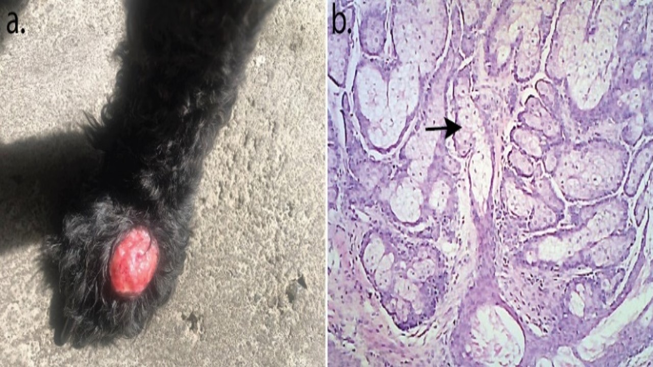
A 14-year-old black male Poodle was brought to the Animal Teaching Hospital, Udayana University by its owner with clinical signs of frequent licking of its left front paw. Upon examination, red bumps were observed on the left front leg, accompanied by small, round black spots scattered on the dorsal side of the body. Additionally, black nodules were present on the lower eyelids and hind limbs. Surgical intervention was undertaken to excise the tumor mass, with the animal under anesthesia induced by ketamine at 5 mg/kg BW intravenously. The reddish nodule was excised by performing an elliptical incision at the base of tumor. Postoperatively, the animal received an antibacterial injection comprising ceftriaxone and tazobactam at 25 mg/kg BW intramuscularly and antiseptic wound dressing for supportive care. Microscopic examination revealed neoplastic cells arranged into lobules of varying sizes and shapes within the tumor mass. These lobules consisted of differentiated sebocytes and basaloid cells. At the periphery of the neoplastic lobules, the basaloid cells displayed several layers and exhibited invasion with moderate anisocytosis. The mitotic index was no more than ten cells in one field of view. Based on these histopathological features, the tumor was confirmed to be a sebaceous adenoma. After a 10-month follow-up period, there were no signs of tumor recurrence observed.
Amaravathi, M., Murthy, R. V. R., Naik, S. H., Nasreen, A., Srilatha, C., Sujatha, K., & Saibaba, M. (2017). Sebaceous gland adenocarcinoma in a dog. Journal of Livestock Science, 8, 18–20.
Angileri, M., Furlanello, T., & de Lucia, M. (2019). Cryotherapy to treat benign skin tumours in conscious dogs. Veterinary Dermatology, 143, 163.
Azevedo, R. S., Almeida, O. P., Netto, J. N. S., Miranda, A. M. M. A., Santos, T. C. R B., Coletta, R. D., Lopes, M. A., & Pires, F. R. (2009). Comparative clinicopathological study of intraoral sebaceous hyperplasia and sebaceous adenoma. Oral Surgery, Oral Medicine, Oral Pathology, Oral Radiology, and Endodontology, 107, 100–104.
Biller, B., Berg, J., & Garrett, L. (2016). AAHA Oncology Guidelines for Dogs and Cats: Veterinary Practice Guidelines. Journal of American Animal Hospital Association, 52, 181–204.
Cohen, P. R., Kohn, S. R., & Kurzrock, R. (1991). Association of sebaceous gland tumors and internal malignancy: The muir-torre syndrome. The American Journal of Medicine, 90, 606–613.
Costa, F. B., da Silva, K. V. G. C., da Silva L. J., Silva, F. B., dos Santos, B.P., de Mello, J. F., & Ferreira, A. M. R. (2020). Histopathological study of canine sebaceous tumors and their association with PCNA expression by immunohistochemistry. Revista Brasileira de Ciencia Veterinaria, 27, 150–158.
Dong, J., Fan, C., Liu, D., & Li, P. (2021). Diagnosis and analysis of a sebaceous gland tumour of the external acoustic meatus in a Cocker Spaniel dog. Acta Veterinaria Brno, 90, 87–89.
Flux, K. (2017). Sebaceous Neoplasms. Surgical Pathology Clinics, 10, 367–382.
Ginel, P. J., Lucena, R., Millán, Y., González-Medina, S., Guil, S., García-Monterde, J., de los Monteros, A. E., & de las Mulas, J. M. (2010). Expression of oestrogen and progesterone receptors in canine sebaceous gland tumours. Veterinary Dermatology, 21, 297–302.
Graf, R., Pospischil, A., Guscetti, F., Meier, D., Welle, M., & Dettwiler, M. (2018). Cutaneous Tumors in Swiss Dogs: Retrospective Data from the Swiss Canine Cancer Registry, 2008–2013. Veterinary Pathology, 55, 809–820.
Gross, T. L., Ihrke, P. J., Walder, E. J., & Affolter, V. K. (2008). Skin diseases of the dog and cat: clinical and histopathologic diagnosis. Blackwell Publishing Company, pp: 641–654.
Kartikasari, A. M., Dewi, C. M. S., Listyasari, N., Pratama, A. R., & Purnama, M. T. E. (2020). Effect of tadpole serum on thyroid hormones and cytotoxic T-cell activity in Wistar rats: a model of skin cancer. Indian Veterinary Journal, 97(6), 34–37.
Kok, M. K., Chambers, J. K., Tsuboi, M., Nishimura, R., Tsujimoto, H., Uchida, K., & Nakayama, H. (2019). Retrospective study of canine cutaneous tumors in Japan, 2008–2017. The Journal of Veterinary Medical Science, 81, 1133–1143.
Lazar, A. J. F, Lyle, S., & Calonje, E. (2007). Sebaceous neoplasia and Torre–Muir syndrome. Current Diagnostic Pathology, 13, 301–319.
MacDonald, V., Turek, M., & Argyle, D. (2008). Tumors of the skin and subcutis. Decision making in small animal oncology. Wiley Blackwell, pp: 129–145.
Martins, A. L., Ana Canadas‑Sousa, A., João, R, Mesquita, J. R., Dias‑Pereira, P., Amorim, I., & Gärtner, F. (2022). Retrospective study of canine cutaneous tumors submitted to a diagnostic pathology laboratory in Northern Portugal (2014–2020). Canine Medicine and Genetics, 9, 2.
Moraes, J., Moraes, E., Baretta, D., Zanetti, A., Garrido, E., Miyazato, L., Sevarolli, A., & Moraes. (2009). Cutaneous Tumors in Dogs - A Retrospective Study of Ten Years. Veterinary Note, 15, 59.
Ozyigit, M. O., Akkoç, A., & Yilmaz, R. (2005). Sebaceous gland adenoma in a dog. Turkish Journal of Veterinary and Animal Sciences, 29, 1213–1216.
Pakhrin, B., Kang, M. S., Bae, I. H., Park, M. S., Jee, H., You, M. H., Kim, J. H., Yoon, B., Choi, Y. K., & Kim, D. Y. (2007). Retrospective study of canine cutaneous tumors in Korea. Journal of Veterinary Medical Science, 8, 229–236.
Parmar, J. J., Shah, A. I., Rao, N., Godasara, D. J., & Patel, D. M. (2019). Successful surgical management of sebaceous gland tumors in dogs. The Indian Journal of Veterinary Sciences and Biotechnology, 15, 79–81.
Patel, M. P., Ghodasara, D. J., Jani, P. B., Joshi, B. P., & Dave, C. J. (2019). Incidence and Histopathology of Sebaceous Gland Tumors in Dogs. Indian Journal of Veterinary Sciences & Biotechnology, 14, 29–32.
Pisani, G., Millanta, F., Lorenzi, D., Vannozzi, I., & Poli, A. (2006). Androgen receptor expression in normal, hyperplastic and neoplastic hepatoid glands in the dog. Research in Veterinary Science, 8, 231–236.
Queiroga, F. L., Pérez-Alenza, D., Silvan, G., Pena, L., & Illera, J. C. (2009). Positive correlation of steroid hormones and EGF in canine mammary cancer. Journal of Steroid Biochemistry and Molecular Biology, 115, 9–13.
Rickyawan, N., Hardian, A. B., & Cadiwirya, P. K. (2021). Lipoma Removal Surgery in White-Rumped Shama (Kittacincla Malabarica Macraoura). Jurnal Medik Veteriner, 4(2), 275–280.
Sabattini, S., Bassi, P., & Bettini, G. (2015). Histopathological Findings and Proliferative Activity of Canine Sebaceous Gland Tumours with a Predominant Reserve Cell Population. Journal of Comparative Pathology, 152, 145–152.
Sananmuang, T., Jeeratanyasakul, P., Mankong, K., & Rattanapinyopituk, K. (2016). Canine sebaceous adenoma of external genitalia: Case report. Journal of Applied Animal Science, 9, 51–56.
Scott, D. W., & Anderson, W. I. (1990). Canine sebaceous gland tumors: a retrospective analysis of 172 cases. Canine Practice, 15, 19–27.
Sewoyo, P. S., & Nainggolan, W. M. (2023). Sebaceous Adenoma Case in a Golden Retriever Dog. Journal of Applied Veterinary Science and Technology, 4, 122–126.
Simkus, D., Pockevicius, A., Maciulskis, P., Simkienė, V., & Zorgevica-Pockevic, L. (2015). Pathomorphological analysis of the most common canine skin and mammary tumors. Veterinarija Ir Zootechnika, 69, 63–70.
Singh, R. S., Grayson, W., & Redston, M. (2008). Site and tumor type predicts DNA mismatch repair status in cutaneous sebaceous neoplasia. The American Journal of Surgical Pathology, 32, 936–942.
Todorova, I. (2006). Prevalence and etiology of the most common malignant tumors in dogs and cats. Bulgarian Journal of Veterinary Medicine, 9, 85–98.
Tozon, N., Kodre, V., Juntes, P., Sersa, G., & Cemazar M. (2010). Electrochemotherapy is highly effective for the treatment of canine perianal hepatoid adenoma epithelioma. Acta Veterinaria, 60, 285–302.
Tozon, N., Kodre, V., Sersa, G., & Cemazar, M. (2005). Effective Treatment of Perianal Tumors in Dogs with Electrochemotherapy. Anticancer Research, 25, 839–846.
Triana, N. M., Wilujeng, E., Putri, M. W. H., Yuda, D. M. P., Hardiono, A. L., Purnama, M. T. E., & Fikri, F. (2020). Antiproliferation effects of Glycine max Linn ethanolic extract on induced mammary gland carcinoma in albino rats. IOP Conference Series: Earth and Environmental Science, 441(1), 012103.
Vail, M. D., & Withrow, S. J. (2007). Tumors of the skin and subcutaneous tissues. In: Small Animal Clinical Oncology. 4th edition. Saunders, pp: 375–401.
Villamil, J. A., Henry, C. J., Bryan, J. N., Ellersieck, M., Schultz, L., Tyler, J. W., & Hahn, A. W. (2011). Identification of the most common cutaneous neoplasms in dogs and evaluation of breed and age distributions for selected neo‑ plasms. Journal of the American Veterinary Medical Association, 239, 960.
Warland, J., & Dobson, J. (2011). Canine and feline skin tumors. Veterinary Focus, 21, 34–41.
Yoon, J., & Park, J. (2016). Immunohistochemical characterization of sebaceous epithelioma in two dogs. Iranian Journal of Veterinary Research, 17, 134.
Copyright (c) 2024 Ida Bagus Oka Winaya, Anak Agung Ayu Mirah Adi, Luh Made Sudimartini, I Made Merdana, Putu Henrywaesa Sudipa, I Gusti Agung Gde Putra Pemayun, Palagan Senopati Sewoyo

This work is licensed under a Creative Commons Attribution-NonCommercial-ShareAlike 4.0 International License.
Authors who publish in this journal agree to the following terms:
1. The journal allows the author to hold the copyright of the article without restrictions;
2. The journal allows the author(s) to retain publishing rights without restrictions;
3. The legal formal aspect of journal publication accessibility refers to Creative Commons Attribution-NonCommercial-ShareAlike 4.0 International License (CC BY-NC-SA).


11.jpg)


