The Detection and Analysis of Factors Associated with the Incidence of Lumpy Skin Disease in Cattle Transhipped at Merak Port, Indonesia

Downloads
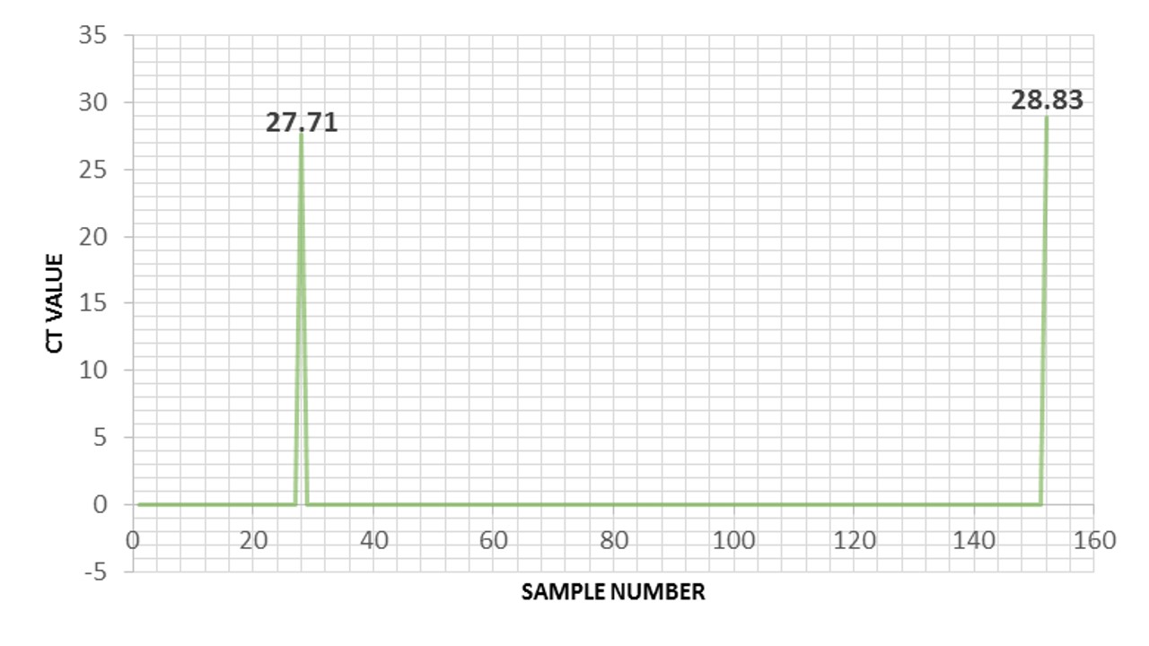
Lumpy Skin Disease (LSD) is caused by the Lumpy Skin Disease Virus (LSDV) that affects cattle and buffalo. The symptoms include the development of lumps or nodules on the skin of infected animals. Therefore, this study aimed to detect the presence of LSD and determine factors associated with the incidence in cattle transhipped through Merak Port. Samples were collected in the form of oral and nasal swabs, then tested with quantitative real-time PCR (qPCR). The results showed that there were two positive LSD samples confirmed by molecular testing using qPCR. The positive cattle did not show clinical signs or were suspected to be sub-clinically infected, while the Ct values obtained were 27.71 and 28.88. The use of molecular methods with qPCR showed relatively good results for the detection of LSD. Cattle that did not show clinical signs were detected as positive by the test. This is because qPCR can detect viruses more quickly and accurately, even at very low viral load levels. Factors associated with the incidence of LSD in the Merak Port (p < 0.05) were farm origin and biosecurity measures, knowledge of livestock handlers, vaccination status, and disinfection practices.
Aerts, L., Haegeman, A., De Leeuw, I., Philips, W., Van Campe, W., Behaeghel, I., Mostin, L., & De Clercq, K. (2021). Detection of clinical and subclinical lumpy skin disease using ear notch testing and skin biopsies. Microorganisms, 9(10), 2171.
Afzal, M. U., Waseem, H., & Li-Ting, C. (2024). Lumpy skin disease: an encroaching risk in cattle farming. Taiwan Veterinary Journal, 49 (1), 1–11.
Aleksandr K., Olga, B., David, W. B., Pavel, P., Yana, P., Svetlana, K., Alexander, N., Vladimir, R., Dmitriy, L., & Alexander, S. (2020). Non-vector-borne transmission of lumpy skin disease virus. Scientific Report, 10, 7436.
Badan Karantina Pertanian (BARANTAN). (2022). Surat Edaran Nomor 5076/KR.120/K/02/2022 tentang Peningkatan Kewaspadaan Terhadap Kejadian Lumpy Skin Disease. Jakarta: Badan Karantina Pertanian.
Bedekovic, T., Simic, I., Kresic, N., & Lojkic, I. (2017). Detection of lumpy skin disease virus in skin lesions, blood, nasal swabs, and milk following preventive vaccination. Transboundary and Emerging Disease, 65(2), 491–496.
Bianchini, J., Simons, X., Humblet, M. F., & Saegerman, C. (2023). Lumpy Skin Disease: a systematic review of mode of transmission, risk of emergence and risk entry pathway. Viruses, 15(8), 1622.
Bowden, T. R., Babiuk, S. L., Parkyn, G. R., Copps, J. S., & Boyle, D. B. (2008). Capripox virus tissue tropism and shedding: a quantitative study in experimentally infected sheep and goats. Virology, 371, 380–393.
Calistri, P., De Clercq, K., Gubbins, S., Klement, E., Stegeman, A., Cortinas Abrahantes, J., Marojevic, D., Antoniou, S. E., & Broglia, A. (2020). Scientific report on the lumpy skin disease epidemiological report IV: Data collection and analysis. EFSA Journal, 18(2), e06010.
Dietze, K., Moritz, T., Alexandrov, T., Krstevski, K., Schlottau, K., Milovanovic, M., Hoffmann, D., & Hoffmann, B. (2018). Suitability of group-level oral fluid sampling in ruminant populations for lumpy skin disease virus detection. Veterinary Microbiology, 221, 44–48.
Direktorat Jenderal Peternakan Kesehatan Hewan & Australia Indonesia Partnership for Emerging Infectious Disease (Ditjen PKH & AIP-EID). (2014). Pedoman Teknis Surveilans Penyakit Hewan Menular. 1st edition. Ditjen PKH. pp: 29–35.
Direktorat Jenderal Peternakan Kesehatan Hewan (Ditjen PKH). (2022). Rencana Kontingensi Lumpy Skin Disease. 1st edition. Ditjen PKH. pp: 1–35.
Fikri, F., & Purnama, M. T. E. (2020). Biosecurity Application of Small Scale Chicken Abattoir in Sidoarjo, East Java, Indonesia. Systematic Reviews in Pharmacy, 11(6), 226–229.
Gumbe, A. A. F. (2018). Review on lumpy skin disease and its economic impact in Ethiopia. Journal Dairy Veterinary Animal Research, 7(1), 39–46.
Gupta, T., Patial, V., Bali, D., Angaria, S., Sharma, M., & Chahota, R. (2020): A review: Lumpy skin disease and its emergence in India. Veterinary Research Communication, 44(3), 111–118.
Hamdi, J., Boumart, Z., Daouam, S., El Arkam, A., Bamouh, Z., Jazouli, M., Tadlaoui, K.O., Fihri, O.F., Gavrilov, B., & El Harrak, M. (2020). Development and evaluation of an inactivated lumpy skin disease vaccine for cattle. Veterinary Microbiology, 245, 108689.
Hilmiat, N. (2019). Cattle farming systems in arid Sumbawa:Opportunities and barriers to improve productivity and farmers income. Journal of Social Economic Agriculture, 13(2), 143–154.
Horigan, V., Beard, P. M., Roberts, H., Adkin. A., Gale, P., Batten, C. A., & Kelly, L. (2018). Assessing the probability of introduction and transmission of Lumpy skin disease virus within the United Kingdom. Microbial Risk Analysis, 9, 1–10.
Indonesian Quarantine Full Automatic System (IQFAST). (2023). Data Lalu Lintas Media Pembawa HPHK Sapi Balai Karantina Kelas II Cilegon. Badan Karantina Pertanian. Jakarta: Badan Karantina Pertanian.
Keputusan Menteri Pertanian Pertanian (KEPMENTAN). (2009). Penggolongan Jenis-jenis Hama Penyakit Hewan Karantina. Jakarta: Kementerian Pertanian RI.
Kononov, A., Byadovskaya, O., Kononova, S., Yashin, R., Zinyakov, N., Mischenko, V., Perevozchikova, N., & Sprygin, A. (2019). Detection of vaccine-like strains of lumpy skin disease virus in outbreaks in Russia in 2017. Archives Virology, 164, 1575–1585.
Krotov, A., Byadovskaya, O., Shumilova. I., van Schalkwyk, A., & Sprygin, A. (2022). An in-depth bioinformatic analysis of the novel recombinant lumpy skin disease virus strains: from unique patterns to established lineage. BMC Genomics, 23, 396.
Leliso, S. A., Bari, F. D., & Chibssa, T. R. (2021). Molecular Characterization of Lumpy Skin Disease Virus Isolates from Outbreak Cases in Cattle from Sawena District of Bale Zone, Oromia, Ethiopia. Veterinary Medicine International, 8862180.
Lestari, V. S., Rahardja, D. P., Sirajuddin, S., & Altawaha, A. R. (2022). Adopting biosecurity measures in cattle breeding systems in Indonesia. Journal of Animal Feed Research, 12(5), 279–283.
Liang, Z., Yao, K., Wang, S., Yin, J., Ma, X., Yin, X., Wang, X., & Sun, Y. (2022). Understanding the research advances on lumpy skin disease: A comprehensive literature review of experimental evidence. Frontiers Microbiology, 28(13), 1065894.
Molla, W., Frankena, K., Gari, G., Kidane, M., Shegu, D., & de Jong, M. C. M. (2018). Seroprevalence and risk factors of lumpy skin disease in Ethiopia. Preventive Veterinary Medicine, 160, 99–104.
Namazi, F. A., & Khodakaram, T. (2021). Lumpy skin disease, an emerging transboundary viral disease: A review. Veterinary Medical Science, 73, 888–896.
Nugroho, E. P., Setiyono, A., Hadi, U. K., Winarsih, W., & Astuti. (2021). Potency of Nuisance Flies as Vector of Coxiella burnetii on Dairy Farms in Ampel and Mojosongo Districts, Boyolali Regency, Middle Java, Indonesia. Advances in Animal Veterinary Sciences, 9(11), 1781–1790.
Nurjanah, D., & Dharmayanti, N. L. P. I. (2022). Ulasan Lumpy Skin Disease: Penyakit infeksius yang berpotensi mengancam kesehatan sapi di Indonesia. Berita Biologi, 21, 1–17.
Pandey, N., Hopker, A., Prajapati, G., Rahangdale, N., Gore, K., & Sargison, N. (2022). Observations on presumptive lumpy skin disease in native cattle and Asian water buffaloes around the tiger reserves of the central Indian highlands, NZ Veterinary Journal, 70, 101–108.
Paramitadevi, Y., Priadi, C., Rahmatika, I., Rukmana, A., & Moersidik, S. (2023). Integration of water, sanitation, and hygiene program with biosecurity: A One Health approach to reduce the prevalence and exposure of antibiotic-resistant bacteria in the livestock community. International Journal of One Health, 9(2), 181–193.
Purnama, M. T. E., Dewi, W. K., Prayoga, S. F., Triana, N. M., Aji, B. S. P., Fikri, F., & Hamid, I. S. (2019). Preslaughter stress in banyuwangi cattle during transport. Indian Veterinary Journal, 96(12), 50–52.
Ratyotha, K., Prakobwong, S., & Piratae, S. (2022). Lumpy skin disease: A newly emerging disease in Southeast Asia. Veterinary World, 15(12), 2764–2771.
Roche, X., Rozstalnyy, A., Tago Pacheco, D., Pittiglio, C., Kamata, A., Beltran Alcrudo, D., Bisht, K., Karki, S., Kayamori, J., Larfaoui, F., Raizman, E., Von Dobschuetz, S., Dhingra, M., & Sumption, K. (2020). Introduction and spread of lumpy skin disease in South, East and Southeast Asia. 1st edition. FAO. pp: 2–35.
Saegerman, C., Bertagnoli, S., Meyer, G., Ganière, J. P., Caufour, P., De Clercq, K., Jacquiet, P., Fournié, G., Hautefeuille, C., Etore, F., & Casal, J. (2018). Risk of introduction of lumpy skin disease in France by the import of vectors in animal trucks. PLoS One, 13(6), e0198506.
Selim, A., Manaa, E., & Khater, H. (2021). Seroprevalence and risk factors for lumpy skin disease in cattle in Northern Egypt. Journal of Tropical Animal Health Production, 53(3), 350.
Sendow, I., Assadah, N. S., Ratnawati, A., Dharmayanti, N. L. P. I., & Saepulloh, M. (2021). Lumpy Skin Disease: ancaman penyakit emerging bagi status kesehatan hewan nasional. Wartazoa, 31(2), 85–96.
Sevik, M., & Dogan, M. (2017). Epidemiological and molecular studies on lumpy skin disease outbreaks in Turkey during 2014–2015. Transboundary and Emerging Disease, 64(4), 1268–1279.
Shumilova, I., Nesterov, A., Byadovskaya, O., Prutnikov, P., Wallace, D. B., Mokeeva, M., Pronin, V., Kononov, A., Chvala, I., & Sprygin, A. (2022). A recombinant vaccine-like strain of lumpy skin disease virus causes low-level infection of cattle through virus-inoculated feed. Pathogens (Basel, Switzerland). Pathogens, 11(8), 920.
Shumilova, I., Prutnikov, P., Mazloum, A., Krotova, A., Tenitilov, N., Byadovskaya, O., Chvala, I., Prokhvatilova, L., & Sprygin, A. (2024). Subclinical infection caused by a recombinant vaccine-like strain poses high risks of lumpy skin disease virus transmission. Frontiers in Veterinary Science, 11, 1330657.
Sikkema, R. S., & Koopmans, M. P. G. (2021). Preparing for Emerging Zoonotic Viruses. Encyclopedia of Virology, 5, 256–266.
Sistem Informasi Kesehatan Hewan Nasional (iSIKHNAS). (2023). Situasi Penyakit Hewan Nasional. Jakarta: Kementerian Pertanian RI.
Sprygin, A., Babin, Y., Pestova, Y., Kononova, S., Wallace, D. B., Van Schalkwyk, A., Byadovskaya, O., Diev, V., Lozovoy, D., & Kononov, A. (2018). Analysis and insights into recombination signals in lumpy skin disease virus recovered in the field. PLoS One, 13, e0207480.
Susanti, T., Susetya, H., Widayani, P., Fitria, Y., & Pambudi, G. T. (2023). Risk factors, logistic model, and vulnerability mapping of lumpy skin disease in livestock at the farm level in Indragiri Hulu District, Riau Province, Indonesia. Veterinary World, 16(10), 2071–2079.
Suwankitwat, N., Bhakha, K., Molee, L., Songkasupa, T., Puangjinda, K., Chamchoy, T., Arjkumpa, O., Nuansrichay, B., Srisomrun, S., Pongphitcha, P., Lekcharoensuk, P., & Arunvipas, P. (2023). Long-term monitoring of immune response to recombinant lumpy skin disease virus in dairy cattle from small-household farms in western Thailand. Comparative Immunology, Microbiology and Infectious Diseases, 99, 102008.
Tuppurainen, E. S. M., Antoniou, S. E., Tsiamadis, E., Topkaridou, M., Labus, T., Debeljak, Z., Plavsic, B., Miteva, A., Alexandrov, T., Pite, L., Boci, J., Marojevic, D., Kondratenko, V., Atanasov, Z., Murati, B., Acinger‐Rogic, Z., Kohnle, L., Calistri, P., & Broglia, A. (2020). Field observations and experiences gained from the implementation of control measures against lumpy skin disease in South‐East Europe between 2015 and 2017. Preventive Veterinary Medicine, 181, 104600.
Tuppurainen, E. S., Venter, E. H., Shisler, J. L., Gari, G., Mekonnen, G. A., Juleff, N., Lyons, N. A., De Clercq, K., Upton, C., Bowden, T. R., Babiuk, S., & Babiuk, L. A. (2017). Review: Capripoxvirus disease: Current status and opportunities for control. Transboundary and Emerging Disease, 64(3), 729–745.
Yuniarti, W. M., Lukiswanto, B. S., Setiawan, B., Yudaniayanti, I. S., Triakoso, N., Madarina Hisyam, M. A., Hastuti, A. N., & Sudjono, B. S. (2024). Strategic Infectious Diseases in Beef Cattle in Balongpanggang during 2023. Jurnal Medik Veteriner, 7(2), 362–369.
Wolff, J., Kiril, K., Martin, B., & Hoffmann, B. (2020). minimum infective dose of a lumpy skin disease virus field strain from north macedonia janika. Viruses, 12(7), 1–15768.
World Organization of Animal Health (WOAH). (2022a). Lumpy Skin Disease. Paris: WOAH.
World Organization of Animal Health (WOAH). (2022b). Terrestrial Animal Health code Chapter 1.4. Animal Health Surveillance. Paris: WOAH.
Zainuddin, M., Maarif, M. S., Rianic, E., & Noor, S. M. (2019). Water Pollution from the Activity of Large-Ruminant Animal Quarantine Installation (AQI) in Its Receiving Water Body. Tropical Animal Science Journal, 42(1), 68–75.
Zeedan, G. S. G., Mahmoud, A. H., Abdalhamed, A. M., El-Razik, K. A. E. A., Khafagi, M. H., & Zeina, H. A. A. A. (2019). Detection of lumpy skin disease virus in cattle using real-time polymerase chain reaction and serological diagnostic assays in different governorates in Egypt in 2017. Veterinary World, 12(7), 1093–1100.
Copyright (c) 2025 Faizal Rafiq, Sri Murtini, Mujiatun Mujiatun, Arum Kusnila Dewi, Melani Wahyu Adiningsih

This work is licensed under a Creative Commons Attribution-NonCommercial-ShareAlike 4.0 International License.
Authors who publish in this journal agree to the following terms:
1. The journal allows the author to hold the copyright of the article without restrictions;
2. The journal allows the author(s) to retain publishing rights without restrictions;
3. The legal formal aspect of journal publication accessibility refers to Creative Commons Attribution-NonCommercial-ShareAlike 4.0 International License (CC BY-NC-SA).


11.jpg)