Pathological Investigation of Lumpy Skin Disease in Cattle from Sleman, Indonesia

Downloads
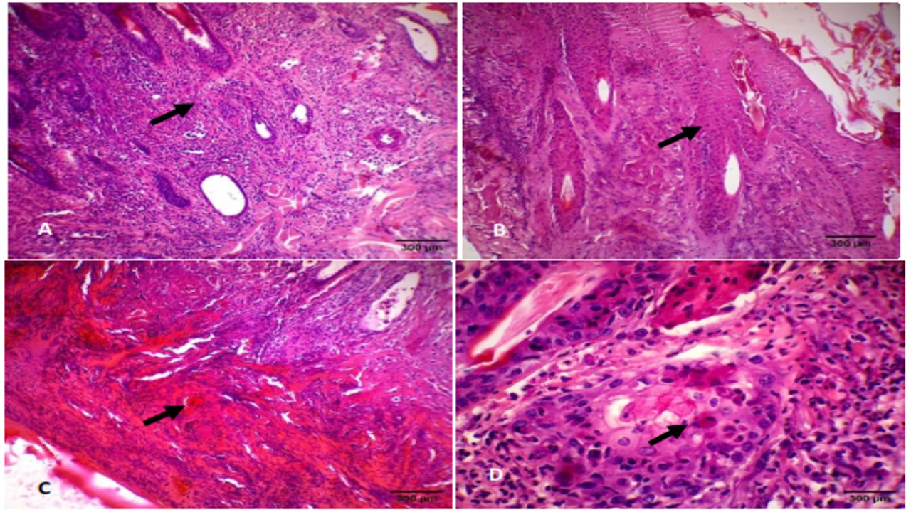
In early 2023, there was an outbreak of lumpy skin disease (LSD) in cattle in Indonesia, with particularly high prevalence in Sleman, Special Region of Yogyakarta, Indonesia. Since then, cases of this disease have been increasing, causing significant economic losses to cattle and buffalo farmers. This study aimed to investigate the pathological changes caused by LSD virus infection in various organs of Sleman cattle. We investigated 15 animals from 10 farms. Skin samples were taken by biopsy. ELISA testing was performed on serum samples. We also performed necropsies on two LSD infected cow carcasses to observe the macroscopic and microscopic effects of the virus. During the necropsies, samples were obtained from skin nodules, skeletal muscle, and internal organs (lung, liver, kidney, lymphatic nodes, spleen, and digestive organs). These were subjected to histopathological examination using hematoxylin and eosin staining. Among the live animals, fever, lethargy, hypersalivation, and lacrimation were the most common clinical signs. Our qualitative descriptive analysis of the pathological changes, clinical signs, and ELISA results showed that LSD infection in cattle causes mild to severe damage to various organs. Our macroscopic examinations found that affected animals had skin nodules of varying sizes over the entire body. We observed mild to severe inflammation and hemorrhage in the internal organs, including the skeletal muscles, spleen, liver, heart, rumen, reticulum, abomasum, and small intestine. Based on this investigation, we conclude that, in addition to its effects on the skin, LSD causes pathological changes in various internal organs.
Abdulqa, H. Y., Rahman, H. S., Dyary, H. O., & Othman, H. H. (2016). Lumpy skin disease. MedPub Journals, 1(4), 1–6.
Abutarbush, S. M. (2017). Lumpy skin disease (knopvelsiekte, pseudo-urticaria, neethling virus disease, exanthema nodularis bovis). In: J. Bayry (Editor), Emerging and re-emerging infectious diseases of livestock. Springer, 12(1), 309–326.
Ali, A. A., Neamat-Allah, A. N., Sheire, H. A. E. M., & Mohamed, R. I. (2021) Prevalence, intensity, and impacts of non-cutaneous lesions of lumpy skin disease among some infected cattle flocks in Nile Delta governorates, Egypt. Comparative Clinical Pathology, 30(4), 693–700.
Arjkumpa, O., Suwannaboon, M., Boonrod, M., Punyawan, I., Liangchaisiri, S., & Laobannue, P. (2022). The first lumpy skin disease outbreak in Thailand (2021): epidemiological features and spatio-temporal analysis. Frontiers in Veterinary Science, 8, 799065.
El-Neweshy, M., El-Shemey, T., & Youssef, S. (2013). Pathologic and immunohistochemical findings of natural lumpy skin disease in Egyptian cattle. Pakistan Veterinary Journal, 33(1), 60–64.
Gharban, H. A., Al-Shaeli, S. J., Al-Fattli, H. H., & Altaee, M. N. (2019). Molecular and histopathological confirmation of clinically diagnosed lumpy skin disease in cattle, Baghdad Province of Iraq. Veterinary World, 12, 1826–1832.
Gupta, T., Patial, V., Bali, D., Angaria, S., Sharma, M., & Chahota, R. (2020). A review: lumpy skin disease and its emergence in India. Veterinary Research Communications, 44(3–4), 111–118.
Ince, O. B. & Turk, T. T. (2019). Analyzing risk factors for lumpy skin disease by a geographic information system (GIS) in Turkey. Journal of the Hellenic Veterinary Medical Society, 70(4), 1797–1804.
Kumar, N., Chander, Y., Kumar, R., Khandelwal, N., Riyesh, T., Chaudhary, K., Shanmugasundaram, K., Kumar, O., Kumar, A., & Gupta, M. K. (2021). Isolation and characterization of lumpy skin disease virus from cattle in India. PLOS ONE, 16(1), e0241022.
Liang, Z., Yao, K., Wang, S., Yin, J., Ma, X., Yin, X., Wang, X., & Sun, Y. (2022). Understanding the research advances on lumpy skin disease: A comprehensive literature review of experimental evidence. Frontiers in Microbiology, 13, 1065894.
Limon, G., Gamawa, A. A., Ahmed, A. I., Lyons, N. A., & Beard, P. M. (2020). Epidemiological characteristics and economic impact of lumpy skin disease, sheeppox and goatpox among subsistence farmers in northeast Nigeria. Frontiers in Veterinary Science, 7, 8.
Manjunathareddy, G. B., Saminathan, M., Sanjeavakumar, L., Rao, S., Dinesh., Dhama, K., Pal Singh, K., & Tripathi, B. N., (2024). Pathological, immunological and molecular epidemiological analysis of lumpy skin disease virus in Indian cattle during a high-mortality epidemic. Veterinary Quarterly, 44(1), 1–22.
Namazi, F., & Tafti, A. K. (2021). Lumpy skin disease, an emerging transboundary viral disease: A review. Veterinary Medicine and Science, 7(3), 888–896.
Nugroho, W., Mardani, H. M., Reichel, M. P., Fitria, Y., Miswati, Y., Febrianto, N., Nuryanto, E., & Apriana, I. (2024). The first outbreak of Lumpy Skin Disease in Indonesia. Tropical Animal Health and Production, 56, 237.
Ochwo, S., VanderWaal, K., Ndekezi, C., Nkamwesiga, J., Munsey, A., Witto, S. G., Nantima, N., Mayanja, F., Okurut, A. R. A., & Atuhaire, D. K. (2020). Molecular detection and phylogenetic analysis of lumpy skin disease virus from outbreaks in Uganda 2017–2018. BMC Veterinary Research, 16(1), 66.
OIE. (2024). Lumpy skin disease. Chapter 3.4.12. OIE Terrestrial Manual, 13, 1–13.
Parvin, R., Chowdhury, H., Islam, M. T., Begum, J. A., Nooruzzaman, M., Globig, A., Dietze, K., Hoffmann, B., & Tuppurainen, E. (2022). Clinical epidemiology, pathology, and molecular investigation of lumpy skin disease outbreaks in Bangladesh during 2020-2021 indicate the re-emergence of an old african strain. Viruses, 14(11), 2529.
Purnama, M. T. E., Dewi, W. K., Prayoga, S. F., Triana, N. M., Aji, B. S. P., Fikri, F., & Hamid, I. S. (2019). Preslaughter stress in banyuwangi cattle during transport. Indian Veterinary Journal, 96(12), 50–52.
Ratyotha, K., Prakobwong, S., & Piratae, S. (2022). Lumpy skin disease: A newly emerging disease in Southeast Asia. Veterinary World, 15(12), 2764–2771.
Razak, A. H. (2023). Kasus LSD pada hewan ternak di Sleman meluas. Harian Jogja. https://jogjapolitan.harianjogja.com/read/2023/04/18/512/1132566/kasus-lsd-di-sleman-mencapai-2124-kasus.
Reddy, G. B. M., Mounica, P. S., Sudeep, N., Vikram, R., Garam, G. B., Lalzampuia, H., Ragulraj, S., Pal, S., Khate, K., Bijalwan, S., Girish, P. S., & Gulati, B. R. (2024). First evidence of lumpy skin disease in mithun (Bos frontalis) in India. Archives of Virology, 169(3), 65.
Sanz-Bernardo, B., Haga, I. R., Wijesiriwardana, N., Hawes, P. C., Simpson, J., Morrison, L. R., MacIntyre, N., Brocchi, E., Atkinson, J., & Haegeman, A. (2020). Lumpy skin disease is characterized by severe multifocal dermatitis with necrotizing fibrinoid vasculitis following experimental infection. Veterinary Pathology, 57(3), 388–396.
Sistem Informasi Kesehatan Hewan Nasional (ISIKHNAS). (2023). Situasi penyakit hewan nasional. ISIKHNAS. https://validation.isikhnas.com
Subarkah, L. (2023). Update sapi LSD Sleman: hingga 9 Februari sudah ada 100 kasus. Harian Jogja. https://jogjapolitan.harianjogja.com/read/2023/02/10/512/1125805/update-sapi-lsd-sleman-hingga-9-februari-sudah-ada-100-kasus.
Tuppurainen, E., Alexandrov, T., & Beltrán-Alcrudo, D. (2017). Lumpy skin disease – A manual for veterinarians. FAO Animal Production and Health Manual No. 20. Rome: Food and Agriculture Organization of the United Nations (FAO), 7–46.
Wick, M. R. (2019). The hematoxylin and eosin stain in anatomic pathology-An often-neglected focus of quality assurance in the laboratory. Seminars in Diagnostic Pathology, 36(5), 303–311.
Copyright (c) 2025 Yuli Purwandari Kristianingrum, Sugi Winarsih, Bambang Sutrisno, Sitarina Widyarini, Sugiyono Sugiyono, Tri Untari

This work is licensed under a Creative Commons Attribution-NonCommercial-ShareAlike 4.0 International License.
Authors who publish in this journal agree to the following terms:
1. The journal allows the author to hold the copyright of the article without restrictions;
2. The journal allows the author(s) to retain publishing rights without restrictions;
3. The legal formal aspect of journal publication accessibility refers to Creative Commons Attribution-NonCommercial-ShareAlike 4.0 International License (CC BY-NC-SA).


11.jpg)