Main Article Content
Abstract
Highlights:
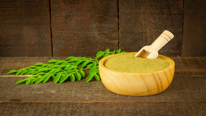
- This study examined the antioxidant flavonoid compounds derived from naturally sourced Moringa oleifera leaves.
- 2. Moringa oleifera leaf extract was able to prevent tissue fibrosis and liver cirrhosis in diabetic rat models through the nonalcoholic fatty liver disease (NAFLD) pathway.
Abstract:
Diabetes mellitus is known as a risk factor for nonalcoholic fatty liver disease (NAFLD) which can progress to nonalcoholic steatohepatitis (NASH) and eventually lead to hepatocellular carcinoma (HCC) through various stages, including necro-inflammatory fibrosis, cirrhosis, and hepatitis. M. oleifera leaves contain flavonoid antioxidants, which inhibit reactive oxygen species (ROS) and oxidative stress in diabetes mellitus. This study aimed to investigate the potential of M. oleifera leaf extract at a dosage of 1,000 mg/kgbw to inhibit liver tissue fibrosis in diabetic rats. This study used a true experimental method with a post-test-only control group design. This study was conducted at the Faculty of Medicine, Universitas Jember, Jember, Indonesia, from November 2021 to January 2022 on 27 male Wistar rats that were divided into three groups of nine rats. The rats were induced with streptozotocin and M. oleifera leaf extract at a dosage of 1,000 mg/kgbw. Masson's trichrome staining and the Meta-analysis of Histological Data in Viral Hepatitis (METAVIR) scoring system were used to measure liver tissue fibrosis. Data were analyzed using the Kruskal-Wallis and Mann-Whitney tests to examine significant differences between groups. The results showed a significant difference in the degree of liver tissue fibrosis between the control and diabetes groups (p=0.00) as well as the diabetes and treatment groups (p=0.003). However, the results did not show any significant differences between the control and treatment groups (p=0.270). These findings suggested that administering M. oleifera leaf extract at a dosage of 1,000 mg/kgbw can inhibit liver tissue fibrosis. In conclusion, this study provides evidence that administering M. oleifera leaf extract can inhibit liver tissue fibrosis in diabetic rats.
Keywords
Article Details
Copyright (c) 2023 Folia Medica Indonesiana

This work is licensed under a Creative Commons Attribution-NonCommercial-ShareAlike 4.0 International License.
-
Folia Medica Indonesiana is a scientific peer-reviewed article which freely available to be accessed, downloaded, and used for research purposes. Folia Medica Indonesiana (p-ISSN: 2541-1012; e-ISSN: 2528-2018) is licensed under a Creative Commons Attribution 4.0 International License. Manuscripts submitted to Folia Medica Indonesiana are published under the terms of the Creative Commons License. The terms of the license are:
Attribution ” You must give appropriate credit, provide a link to the license, and indicate if changes were made. You may do so in any reasonable manner, but not in any way that suggests the licensor endorses you or your use.
NonCommercial ” You may not use the material for commercial purposes.
ShareAlike ” If you remix, transform, or build upon the material, you must distribute your contributions under the same license as the original.
No additional restrictions ” You may not apply legal terms or technological measures that legally restrict others from doing anything the license permits.
You are free to :
Share ” copy and redistribute the material in any medium or format.
Adapt ” remix, transform, and build upon the material.

References
- Adeyemi DO, Ukwenya VO, Obuotor EM, et al (2014). Anti-hepatotoxic activities of Hibiscus sabdariffa L. in animal model of streptozotocin diabetes-induced liver damage. BMC Complementary and Alternative Medicine14,
- doi: 10.1186/1472-6882-14-277.
- Akhlaghi M (2016). Non-alcoholic fatty liver disease: Beneficial effects of flavonoids. Phytotherapy Research 30, 1559–1571. doi: 10.1002/ptr.5667
- American Veterinary Medical Association (2020). AVMA guidelines for the euthanasia of animals. AVMA. Available at: https://www.avma.org/resources-tools/avma-policies/avma-guidelines-euthanasia-animalsAseer KR, Kim SW, Choi M-S, et al (2015). Opposite expression of SPARC between the liver and pancreas in streptozotocin-induced diabetic rats ed. Cheng J-T. PLoS One 10, e0131189. doi: 10.1371/journal.pone.0131189
- Bellentani S (2017). The epidemiology of non-alcoholic fatty liver disease. Liver International 37, 81–84. doi: 10.1111/liv.13299
- Chengxi L, Rentao L, Wei Z (2018). Progress in non-invasive detection of liver fibrosis. Cancer Biology Medicine 15, 124. doi: 10.20892/j.issn.2095-3941.2018.0018
- Choo D, Shin KS, Min JH, et al (2022). Noninvasive assessment of liver fibrosis with ElastPQ in patients with chronic viral hepatitis: Comparison using histopathological findings. Diagnostics 12, 706. doi: 10.3390/diagnostics12030706
- Fulton DJR, Li X, Bordan Z, et al (2019). Galectin-3: A harbinger of reactive oxygen species, fibrosis, and inflammation in pulmonary arterial hypertension. Antioxidant & Redox Signaling 31, 1053–1069. doi: 10.1089/ars.2019.7753
- Furman BL (2021). Streptozotocin"induced diabetic models in mice and rats. Current Protocols. doi: 10.1002/cpz1.78.
- Gheibi S, Kashfi K, Ghasemi A (2017). A practical guide for induction of type-2 diabetes in rat: Incorporating a high-fat diet and streptozotocin. Biomedicine & Pharmacotherapy 95, 605–613. doi: 10.1016/j.biopha.2017.08.098
- Hardianto D (2021). Telaah komprehensif diabetes melitus: Klasifikasi, gejala, diagnosis, pencegahan, dan pengobatan. Jurnal Bioteknologi & amp; Biosains Indonesia 7, 304–317. doi: 10.29122/jbbi.v7i2.4209
- Hazlehurst JM, Woods C, Marjot T, et al. (2016). Non-alcoholic fatty liver disease and diabetes. Metabolism 65, 1096–1108. doi: 10.1016/j.metabol.2016.01.001
- Heydarpour F, Sajadimajd S, Mirzarazi E, et al (2020). Involvement of TGF-β and autophagy pathways in pathogenesis of diabetes: A comprehensive review on biological and pharmacological insights. Frontiers in Pharmacology. doi: 10.3389/fphar.2020.498758.
- Kim J, Kang W, Kang SH, et al (2020). Proline-rich tyrosine kinase 2 mediates transforming growth factor-beta-induced hepatic stellate cell activation and liver fibrosis. Scientific Reports 10, 21018. doi: 10.1038/s41598-020-78056-0
- Leite NC (2014). Non-alcoholic fatty liver disease and diabetes: From physiopathological interplay to diagnosis and treatment. World Journal of Gastroenterology 20, 8377. doi: 10.3748/wjg.v20.i26.8377
- Lin M, Zhang J, Chen X (2018). Bioactive flavonoids in Moringa oleifera and their health-promoting properties. Journal of Function Foods 47, 469–479. doi: 10.1016/j.jff.2018.06.011
- Minister of Health of the Republic of Indonesia (2018). Hari Diabetes Sedunia tahun 2018. Minist Heal Repub Indones. Available at: https://p2ptm.kemkes.go.id/tag/hari-diabetes-sedunia-tahun-2018.
- Mitra S, De A, Chowdhury A (2020). Epidemiology of non-alcoholic and alcoholic fatty liver diseases. Translational Gastroenterology Hepatology 5, 16. doi: 10.21037/tgh.2019.09.08
- Moreli JB, Santos JH, Rocha CR, et al (2014). DNA damage and its cellular response in mother and fetus exposed to hyperglycemic environment. Biomed Research International 2014, 1–9. doi: 10.1155/2014/676758
- Nasution F, Andilala A, Siregar AA (2021). Faktor risiko kejadian diabetes melitus. Jurnal Ilmu Kesehatan 9, 94. doi: 10.32831/jik.v9i2.304
- Nortjie E, Basitere M, Moyo D, et al (2022). Extraction methods, quantitative and qualitative phytochemical screening of medicinal plants for antimicrobial textiles: A review. Plants 11, 2011. doi: 10.3390/plants11152011
- Ramadan MA, Pramaningtyas MD, Lusiantari R (2022). Derajat fibrosis dan skor nafld pada hepar tikus diabetes mellitus tipe 2 remaja. Jurnal Kedokteran dan Kesehatan: Publikasi Ilmiah Fakultas Kedokteran Universitas Sriwijaya 9, 83–90. doi: 10.32539/JKK.V9I1.151315
- Safithri F (2018). Mekanisme regenerasi hati secara endogen pada fibrosis hati. MAGNA MEDICA: Berkala Ilmiah Kedokteran dan Kesehatan 2, 9. doi: 10.26714/magnamed.2.4.2018.9-26
- Salih ND, Kumar GH, Noah RM, et al (2014). The effect of streptozotocin induced diabetes mellitus on liver activity in mice. Global Journal on Advances in Pure & Applied Sciences 3, 67–75. Available at: https://www.researchgate.net/publication/324279523_The_effect_of_streptozotocin_induced_diabetes_mellitus_on_liver_activity_in_mice
- Saputra AD (2018). Studi tingkat kecelakaan lalu lintas jalan di Indonesia berdasarkan data KNKT (Komite Nasional Keselamatan Transportasi) dari tahun 2007-2016. Warta Penelitian Perhubungan 29, 179. doi: 10.25104/warlit.v29i2.557
- Tukiran, Miranti MG, Dianawati I, et al (2020). Aktivitas antioksidan ekstrak daun kelor (Moringa oleifera Lam.) dan buah bit (Beta vulgaris L.) sebagai bahan tanbahan minuman suplemen. Jurnal Kimia Riset 5, 113. doi: 10.20473/jkr.v5i2.22518
- van de Vlekkert D, Machado E, D'Azzo A (2020). Analysis of generalized fibrosis in mouse tissue sections with Masson's Trichrome staining. BIO-PROTOCOL. doi: 10.21769/BioProtoc.3629 World Health Organization (2020). Diabetes. WHO. Available at: https://www.who.int/news/item/09-12-2020-who-reveals-leading-causes-of-death-and-disability-worldwide-2000-2019.
- Xia M-F, Bian H, Gao X (2019). NAFLD and diabetes: Two sides of the same coin? rationale for gene-based personalized NAFLD treatment. Frontiers in Pharmacology. doi: 10.3389/fphar.2019.00877
- Yang L, Li L-C, Lamaoqiezhong, et al (2019). The contributions of mesoderm-derived cells in liver development. Seminars in Cell & Developmental Biology 92, 63–76. doi: 10.1016/j.semcdb.2018.09.003
- Zhao Y-L, Zhu R-T, Sun Y-L (2016). Epithelial-mesenchymal transition in liver fibrosis. Biomedical Reports 4, 269–274. doi:
- 3892/br.2016.578
References
Adeyemi DO, Ukwenya VO, Obuotor EM, et al (2014). Anti-hepatotoxic activities of Hibiscus sabdariffa L. in animal model of streptozotocin diabetes-induced liver damage. BMC Complementary and Alternative Medicine14,
doi: 10.1186/1472-6882-14-277.
Akhlaghi M (2016). Non-alcoholic fatty liver disease: Beneficial effects of flavonoids. Phytotherapy Research 30, 1559–1571. doi: 10.1002/ptr.5667
American Veterinary Medical Association (2020). AVMA guidelines for the euthanasia of animals. AVMA. Available at: https://www.avma.org/resources-tools/avma-policies/avma-guidelines-euthanasia-animalsAseer KR, Kim SW, Choi M-S, et al (2015). Opposite expression of SPARC between the liver and pancreas in streptozotocin-induced diabetic rats ed. Cheng J-T. PLoS One 10, e0131189. doi: 10.1371/journal.pone.0131189
Bellentani S (2017). The epidemiology of non-alcoholic fatty liver disease. Liver International 37, 81–84. doi: 10.1111/liv.13299
Chengxi L, Rentao L, Wei Z (2018). Progress in non-invasive detection of liver fibrosis. Cancer Biology Medicine 15, 124. doi: 10.20892/j.issn.2095-3941.2018.0018
Choo D, Shin KS, Min JH, et al (2022). Noninvasive assessment of liver fibrosis with ElastPQ in patients with chronic viral hepatitis: Comparison using histopathological findings. Diagnostics 12, 706. doi: 10.3390/diagnostics12030706
Fulton DJR, Li X, Bordan Z, et al (2019). Galectin-3: A harbinger of reactive oxygen species, fibrosis, and inflammation in pulmonary arterial hypertension. Antioxidant & Redox Signaling 31, 1053–1069. doi: 10.1089/ars.2019.7753
Furman BL (2021). Streptozotocin"induced diabetic models in mice and rats. Current Protocols. doi: 10.1002/cpz1.78.
Gheibi S, Kashfi K, Ghasemi A (2017). A practical guide for induction of type-2 diabetes in rat: Incorporating a high-fat diet and streptozotocin. Biomedicine & Pharmacotherapy 95, 605–613. doi: 10.1016/j.biopha.2017.08.098
Hardianto D (2021). Telaah komprehensif diabetes melitus: Klasifikasi, gejala, diagnosis, pencegahan, dan pengobatan. Jurnal Bioteknologi & amp; Biosains Indonesia 7, 304–317. doi: 10.29122/jbbi.v7i2.4209
Hazlehurst JM, Woods C, Marjot T, et al. (2016). Non-alcoholic fatty liver disease and diabetes. Metabolism 65, 1096–1108. doi: 10.1016/j.metabol.2016.01.001
Heydarpour F, Sajadimajd S, Mirzarazi E, et al (2020). Involvement of TGF-β and autophagy pathways in pathogenesis of diabetes: A comprehensive review on biological and pharmacological insights. Frontiers in Pharmacology. doi: 10.3389/fphar.2020.498758.
Kim J, Kang W, Kang SH, et al (2020). Proline-rich tyrosine kinase 2 mediates transforming growth factor-beta-induced hepatic stellate cell activation and liver fibrosis. Scientific Reports 10, 21018. doi: 10.1038/s41598-020-78056-0
Leite NC (2014). Non-alcoholic fatty liver disease and diabetes: From physiopathological interplay to diagnosis and treatment. World Journal of Gastroenterology 20, 8377. doi: 10.3748/wjg.v20.i26.8377
Lin M, Zhang J, Chen X (2018). Bioactive flavonoids in Moringa oleifera and their health-promoting properties. Journal of Function Foods 47, 469–479. doi: 10.1016/j.jff.2018.06.011
Minister of Health of the Republic of Indonesia (2018). Hari Diabetes Sedunia tahun 2018. Minist Heal Repub Indones. Available at: https://p2ptm.kemkes.go.id/tag/hari-diabetes-sedunia-tahun-2018.
Mitra S, De A, Chowdhury A (2020). Epidemiology of non-alcoholic and alcoholic fatty liver diseases. Translational Gastroenterology Hepatology 5, 16. doi: 10.21037/tgh.2019.09.08
Moreli JB, Santos JH, Rocha CR, et al (2014). DNA damage and its cellular response in mother and fetus exposed to hyperglycemic environment. Biomed Research International 2014, 1–9. doi: 10.1155/2014/676758
Nasution F, Andilala A, Siregar AA (2021). Faktor risiko kejadian diabetes melitus. Jurnal Ilmu Kesehatan 9, 94. doi: 10.32831/jik.v9i2.304
Nortjie E, Basitere M, Moyo D, et al (2022). Extraction methods, quantitative and qualitative phytochemical screening of medicinal plants for antimicrobial textiles: A review. Plants 11, 2011. doi: 10.3390/plants11152011
Ramadan MA, Pramaningtyas MD, Lusiantari R (2022). Derajat fibrosis dan skor nafld pada hepar tikus diabetes mellitus tipe 2 remaja. Jurnal Kedokteran dan Kesehatan: Publikasi Ilmiah Fakultas Kedokteran Universitas Sriwijaya 9, 83–90. doi: 10.32539/JKK.V9I1.151315
Safithri F (2018). Mekanisme regenerasi hati secara endogen pada fibrosis hati. MAGNA MEDICA: Berkala Ilmiah Kedokteran dan Kesehatan 2, 9. doi: 10.26714/magnamed.2.4.2018.9-26
Salih ND, Kumar GH, Noah RM, et al (2014). The effect of streptozotocin induced diabetes mellitus on liver activity in mice. Global Journal on Advances in Pure & Applied Sciences 3, 67–75. Available at: https://www.researchgate.net/publication/324279523_The_effect_of_streptozotocin_induced_diabetes_mellitus_on_liver_activity_in_mice
Saputra AD (2018). Studi tingkat kecelakaan lalu lintas jalan di Indonesia berdasarkan data KNKT (Komite Nasional Keselamatan Transportasi) dari tahun 2007-2016. Warta Penelitian Perhubungan 29, 179. doi: 10.25104/warlit.v29i2.557
Tukiran, Miranti MG, Dianawati I, et al (2020). Aktivitas antioksidan ekstrak daun kelor (Moringa oleifera Lam.) dan buah bit (Beta vulgaris L.) sebagai bahan tanbahan minuman suplemen. Jurnal Kimia Riset 5, 113. doi: 10.20473/jkr.v5i2.22518
van de Vlekkert D, Machado E, D'Azzo A (2020). Analysis of generalized fibrosis in mouse tissue sections with Masson's Trichrome staining. BIO-PROTOCOL. doi: 10.21769/BioProtoc.3629 World Health Organization (2020). Diabetes. WHO. Available at: https://www.who.int/news/item/09-12-2020-who-reveals-leading-causes-of-death-and-disability-worldwide-2000-2019.
Xia M-F, Bian H, Gao X (2019). NAFLD and diabetes: Two sides of the same coin? rationale for gene-based personalized NAFLD treatment. Frontiers in Pharmacology. doi: 10.3389/fphar.2019.00877
Yang L, Li L-C, Lamaoqiezhong, et al (2019). The contributions of mesoderm-derived cells in liver development. Seminars in Cell & Developmental Biology 92, 63–76. doi: 10.1016/j.semcdb.2018.09.003
Zhao Y-L, Zhu R-T, Sun Y-L (2016). Epithelial-mesenchymal transition in liver fibrosis. Biomedical Reports 4, 269–274. doi:
3892/br.2016.578

