

Article Sidebar
Main Article Content
Abstract
Highlights:
- This original research report on the differences in disease severity and blood test results between unvaccinated and vaccinated COVID-19 patients is quite novel, as the COVID-19 pandemic was still happening when this study was carried out.
- This original research report offers information on the advantages of the COVID-19 vaccines and provides data that the public may use as scientific evidence to counter hoaxes.
Abstract
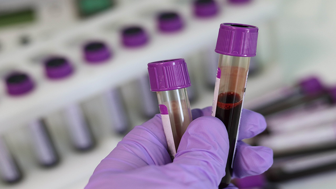
The COVID-19 pandemic caused by SARS-CoV-2 is an unprecedented event in human history. Vaccines are a safe, long-term solution for addressing the COVID-19 pandemic. This study aimed to investigate the differences in disease severity and blood test results between unvaccinated and vaccinated COVID-19 patients. This study used an analytical observational method with purposive sampling. A total of 90 COVID-19 patients at Bhayangkara Hospital, Denpasar, Indonesia, were divided into three groups: unvaccinated group (V0), two-dose vaccinated group (Vp), and three-dose vaccinated group (Vb). Primary data were collected from July to December 2022, while secondary data were collected from January 2021 to June 2022. The data were analyzed using the Kruskal-Wallis test followed by the Mann-Whitney test, as well as one-way ANOVA test followed by Tukey's honestly significant difference (HSD) test with a confidence interval (CI) of 95% and α of 5%. The results revealed significant differences in disease severity (p<0.001). V0 had a higher percentage of severe (36.7%) and critical (6.7%) symptoms than Vp (severe=10.0%; critical, n=0) and Vb (severe and critical, n=0). The follow-up tests revealed significant differences in disease severity between V0 and Vp (p<0.001), V0 and Vb (p<0.001), as well as Vp and Vb (p=0.001). Blood test results revealed significant differences in lymphocytes (p=0.005), monocytes (p<0.001), monocyte-to-lymphocyte ratio (MLR) (p<0.001), and eosinophils (p=0.037). The follow-up tests revealed significant differences in these four indicators between V0 and Vb, in all parameters except for lymphocytes between V0 and Vp, and in lymphocytes only between Vp vs Vb. In conclusion, unvaccinated patients had a higher percentage of severe and critical symptoms than vaccinated patients. The blood test results revealed significant differences in lymphocytes, monocytes, MLR, and eosinophils. Unvaccinated patients had lower lymphocyte counts, higher MLR levels, and higher monocyte counts than vaccinated patients.
Keywords
Article Details
Copyright (c) 2023 Folia Medica Indonesiana

This work is licensed under a Creative Commons Attribution-NonCommercial-ShareAlike 4.0 International License.
-
Folia Medica Indonesiana is a scientific peer-reviewed article which freely available to be accessed, downloaded, and used for research purposes. Folia Medica Indonesiana (p-ISSN: 2541-1012; e-ISSN: 2528-2018) is licensed under a Creative Commons Attribution 4.0 International License. Manuscripts submitted to Folia Medica Indonesiana are published under the terms of the Creative Commons License. The terms of the license are:
Attribution ” You must give appropriate credit, provide a link to the license, and indicate if changes were made. You may do so in any reasonable manner, but not in any way that suggests the licensor endorses you or your use.
NonCommercial ” You may not use the material for commercial purposes.
ShareAlike ” If you remix, transform, or build upon the material, you must distribute your contributions under the same license as the original.
No additional restrictions ” You may not apply legal terms or technological measures that legally restrict others from doing anything the license permits.
You are free to :
Share ” copy and redistribute the material in any medium or format.
Adapt ” remix, transform, and build upon the material.

References
- Antonelli M, Penfold RS, Merino J, et al (2022). Risk factors and disease profile of post-vaccination SARS-CoV-2 infection in UK users of the COVID Symptom Study app: a prospective, community-based, nested, case-control study. The Lancet Infectious Diseases 22, 43–55. doi: 10.1016/S1473-3099(21)00460-6.
- Bakasis A-D, Mavragani CP, Voulgari P V., et al (2022). COVID-19: Clinical features and outcomes in unvaccinated 2-dose and 3-dose vaccinated against SARS-CoV-2 patients with systemic autoimmune and autoinflammatory rheumatic diseases. Journal of Autoimmunity 131, 102846. doi: 10.1016/j.jaut.2022.102846.
- Citu C, Gorun F, Motoc A, et al (2022). The predictive role of NLR, d-NLR, MLR, and SIRI in COVID-19 mortality. Diagnostics 12, 122. doi: 10.3390/diagnostics12010122.
- Cleophas TJ, Zwinderman AH (2016). Non-parametric tests for three or more samples (Friedman and Kruskal-Wallis). Clinical Data Analysis on a Pocket Calculator, 193–7. Springer International Publishing, Cham. doi: 10.1007/978-3-319-27104-0_34.
- Diao B, Wang C, Tan Y, et al (2020). Reduction and functional exhaustion of T cells in patients with coronavirus disease 2019 (COVID-19). Frontiers in Immunology. doi: 10.3389/fimmu.2020.00827.
- Erdogan A, Can FE, Gönüllü H (2021). Evaluation of the prognostic role of NLR, LMR, PLR, and LCR ratio in COVID"19 patients. Journal of Medical Virology 93, 5555–5559. doi: 10.1002/jmv.27097.
- Ertekin B, Yortanlı M, Özelbaykal O, et al (2021). The relationship between routine blood parameters and the prognosis of COVID-19 patients in the emergency department ed. Kam CW. Emergency Medicine International 2021, 1–7. doi: 10.1155/2021/7489675.
- Hanafi M, E Linawati, W Soewondo (2021). Thorax imaging of vaccinated and non-vaccinated Covid-19 patients, how are they different? GSC Advanced Research and Reviews 9, 185–189. doi: 10.30574/gscarr.2021.9.1.0253.
- Hartono CE, Tresia L, Nathania VA, et al (2022). The impact of hoax on COVID-19 vaccination Indonesia. Academy of Education Journal 13, 210–223. doi: 10.47200/aoej.v13i2.1005.
- Hernaningsih Y (2021). Aspek Laboratorium Covid-19. Airlangga University Press, Surabaya.
- Huang I, Pranata R (2020). Lymphopenia in severe coronavirus disease-2019 (COVID-19): systematic review and meta-analysis. Journal of Intensive Care 8, 36. doi: 10.1186/s40560-020-00453-4.
- Islas-Vazquez L, Cruz-Aguilar M, Velazquez-Soto H, et al (2022). Effector-memory B-lymphocytes and follicular helper T-lymphocytes as central players in the immune response in vaccinated and nonvaccinated populations against SARS-CoV-2. Vaccines 10, 1761. doi: 10.3390/vaccines10101761.
- Lee SW (2022). Methods for testing statistical differences between groups in medical research: statistical standard and guideline of Life Cycle Committee. Life Cycle. doi: 10.54724/lc.2022.e1.
- Lindsley AW, Schwartz JT, Rothenberg ME (2020). Eosinophil responses during COVID-19 infections and coronavirus vaccination. Journal of Allergy and Clinical Immunology 146, 1–7. doi: 10.1016/j.jaci.2020.04.021.
- Mao J, Dai R, Du R-C, et al (2021). Hematologic changes predict clinical outcome in recovered patients with COVID-19. Annals of Hematology 100, 675–689. doi: 10.1007/s00277-021-04426-x.
- Mazzoni A, Salvati L, Maggi L, et al (2020). Impaired immune cell cytotoxicity in severe COVID-19 is IL-6 dependent. Journal of Clinical Investigation 130, 4694–4703. doi: 10.1172/JCI138554.
- Ok F, Erdogan O, Durmus E, et al. (2021). Predictive values of blood urea nitrogen/creatinine ratio and other routine blood parameters on disease severity and survival of COVID"19 patients. Journal of Medical Virology 93, 786–793. doi: 10.1002/jmv.26300.
- Ouyang Y, Yin J, Wang W, et al (2020). Downregulated gene expression spectrum and immune responses changed during the disease progression in patients with COVID-19. Clinical Infectious Diseases 71, 2052–2060. doi: 10.1093/cid/ciaa462.
- Peng J, Qi D, Yuan G, et al. (2020). Diagnostic value of peripheral hematologic markers for coronavirus disease 2019 (COVID"19): A multicenter, cross"sectional study. Journal of Clinical Laboratory Analysis. doi: 10.1002/jcla.23475.
- Porto LC, Costa CH, Nunes AS, et al. (2022). Clinical and laboratory characteristics in outpatient diagnosis of COVID-19 in healthcare professionals in Rio de Janeiro, Brazil. Journal of Clinical Pathology 75, 185–192. doi: 10.1136/jclinpath-2020-206797.
- Saija VJE (2021). Covid-19 vaccination: Rights or obligations? SASI 27, 430. doi: 10.47268/sasi.v27i4.683.
- Sun S, Cai X, Wang H, et al. (2020). Abnormalities of peripheral blood system in patients with COVID-19 in Wenzhou, China. Clinica Chimica Acta 507, 174–180. doi: 10.1016/j.cca.2020. 04.024.
- Tanni F, Akker E, Zaman MM, et al (2020). Eosinopenia and COVID-19. Journal of Osteopathic Medicine 120, 504–508. doi: 10.7556/jaoa.2020.091.
- Tavakolpour S, Rakhshandehroo T, Wei EX, et al (2020). Lymphopenia during the COVID-19 infection: What it shows and what can be learned. Immunology Letters 225, 31–32. doi: 10.1016/j.imlet.2020.06.013.
- Zhang J, Dong X, Cao Y, et al (2020). Clinical characteristics of 140 patients infected with SARS"CoV"2 in Wuhan, China. Allergy 75, 1730–1741. doi: 10.1111/all.14238.
- Zhao Q, Meng M, Kumar R, et al (2020). Lymphopenia is associated with severe coronavirus disease 2019 (COVID-19) infections: A systemic review and meta-analysis. International Journal of Infectious Diseases 96, 131–135. doi: 10.1016/j.ijid.2020.04.086.
- Zhou Y, Fu B, Zheng X, et al. (2020). Pathogenic T-cells and inflammatory monocytes incite inflammatory storms in severe COVID-19 patients. National Science Review 7, 998–1002. doi: 10.1093/nsr/nwaa041.
References
Antonelli M, Penfold RS, Merino J, et al (2022). Risk factors and disease profile of post-vaccination SARS-CoV-2 infection in UK users of the COVID Symptom Study app: a prospective, community-based, nested, case-control study. The Lancet Infectious Diseases 22, 43–55. doi: 10.1016/S1473-3099(21)00460-6.
Bakasis A-D, Mavragani CP, Voulgari P V., et al (2022). COVID-19: Clinical features and outcomes in unvaccinated 2-dose and 3-dose vaccinated against SARS-CoV-2 patients with systemic autoimmune and autoinflammatory rheumatic diseases. Journal of Autoimmunity 131, 102846. doi: 10.1016/j.jaut.2022.102846.
Citu C, Gorun F, Motoc A, et al (2022). The predictive role of NLR, d-NLR, MLR, and SIRI in COVID-19 mortality. Diagnostics 12, 122. doi: 10.3390/diagnostics12010122.
Cleophas TJ, Zwinderman AH (2016). Non-parametric tests for three or more samples (Friedman and Kruskal-Wallis). Clinical Data Analysis on a Pocket Calculator, 193–7. Springer International Publishing, Cham. doi: 10.1007/978-3-319-27104-0_34.
Diao B, Wang C, Tan Y, et al (2020). Reduction and functional exhaustion of T cells in patients with coronavirus disease 2019 (COVID-19). Frontiers in Immunology. doi: 10.3389/fimmu.2020.00827.
Erdogan A, Can FE, Gönüllü H (2021). Evaluation of the prognostic role of NLR, LMR, PLR, and LCR ratio in COVID"19 patients. Journal of Medical Virology 93, 5555–5559. doi: 10.1002/jmv.27097.
Ertekin B, Yortanlı M, Özelbaykal O, et al (2021). The relationship between routine blood parameters and the prognosis of COVID-19 patients in the emergency department ed. Kam CW. Emergency Medicine International 2021, 1–7. doi: 10.1155/2021/7489675.
Hanafi M, E Linawati, W Soewondo (2021). Thorax imaging of vaccinated and non-vaccinated Covid-19 patients, how are they different? GSC Advanced Research and Reviews 9, 185–189. doi: 10.30574/gscarr.2021.9.1.0253.
Hartono CE, Tresia L, Nathania VA, et al (2022). The impact of hoax on COVID-19 vaccination Indonesia. Academy of Education Journal 13, 210–223. doi: 10.47200/aoej.v13i2.1005.
Hernaningsih Y (2021). Aspek Laboratorium Covid-19. Airlangga University Press, Surabaya.
Huang I, Pranata R (2020). Lymphopenia in severe coronavirus disease-2019 (COVID-19): systematic review and meta-analysis. Journal of Intensive Care 8, 36. doi: 10.1186/s40560-020-00453-4.
Islas-Vazquez L, Cruz-Aguilar M, Velazquez-Soto H, et al (2022). Effector-memory B-lymphocytes and follicular helper T-lymphocytes as central players in the immune response in vaccinated and nonvaccinated populations against SARS-CoV-2. Vaccines 10, 1761. doi: 10.3390/vaccines10101761.
Lee SW (2022). Methods for testing statistical differences between groups in medical research: statistical standard and guideline of Life Cycle Committee. Life Cycle. doi: 10.54724/lc.2022.e1.
Lindsley AW, Schwartz JT, Rothenberg ME (2020). Eosinophil responses during COVID-19 infections and coronavirus vaccination. Journal of Allergy and Clinical Immunology 146, 1–7. doi: 10.1016/j.jaci.2020.04.021.
Mao J, Dai R, Du R-C, et al (2021). Hematologic changes predict clinical outcome in recovered patients with COVID-19. Annals of Hematology 100, 675–689. doi: 10.1007/s00277-021-04426-x.
Mazzoni A, Salvati L, Maggi L, et al (2020). Impaired immune cell cytotoxicity in severe COVID-19 is IL-6 dependent. Journal of Clinical Investigation 130, 4694–4703. doi: 10.1172/JCI138554.
Ok F, Erdogan O, Durmus E, et al. (2021). Predictive values of blood urea nitrogen/creatinine ratio and other routine blood parameters on disease severity and survival of COVID"19 patients. Journal of Medical Virology 93, 786–793. doi: 10.1002/jmv.26300.
Ouyang Y, Yin J, Wang W, et al (2020). Downregulated gene expression spectrum and immune responses changed during the disease progression in patients with COVID-19. Clinical Infectious Diseases 71, 2052–2060. doi: 10.1093/cid/ciaa462.
Peng J, Qi D, Yuan G, et al. (2020). Diagnostic value of peripheral hematologic markers for coronavirus disease 2019 (COVID"19): A multicenter, cross"sectional study. Journal of Clinical Laboratory Analysis. doi: 10.1002/jcla.23475.
Porto LC, Costa CH, Nunes AS, et al. (2022). Clinical and laboratory characteristics in outpatient diagnosis of COVID-19 in healthcare professionals in Rio de Janeiro, Brazil. Journal of Clinical Pathology 75, 185–192. doi: 10.1136/jclinpath-2020-206797.
Saija VJE (2021). Covid-19 vaccination: Rights or obligations? SASI 27, 430. doi: 10.47268/sasi.v27i4.683.
Sun S, Cai X, Wang H, et al. (2020). Abnormalities of peripheral blood system in patients with COVID-19 in Wenzhou, China. Clinica Chimica Acta 507, 174–180. doi: 10.1016/j.cca.2020. 04.024.
Tanni F, Akker E, Zaman MM, et al (2020). Eosinopenia and COVID-19. Journal of Osteopathic Medicine 120, 504–508. doi: 10.7556/jaoa.2020.091.
Tavakolpour S, Rakhshandehroo T, Wei EX, et al (2020). Lymphopenia during the COVID-19 infection: What it shows and what can be learned. Immunology Letters 225, 31–32. doi: 10.1016/j.imlet.2020.06.013.
Zhang J, Dong X, Cao Y, et al (2020). Clinical characteristics of 140 patients infected with SARS"CoV"2 in Wuhan, China. Allergy 75, 1730–1741. doi: 10.1111/all.14238.
Zhao Q, Meng M, Kumar R, et al (2020). Lymphopenia is associated with severe coronavirus disease 2019 (COVID-19) infections: A systemic review and meta-analysis. International Journal of Infectious Diseases 96, 131–135. doi: 10.1016/j.ijid.2020.04.086.
Zhou Y, Fu B, Zheng X, et al. (2020). Pathogenic T-cells and inflammatory monocytes incite inflammatory storms in severe COVID-19 patients. National Science Review 7, 998–1002. doi: 10.1093/nsr/nwaa041.