Ultrasonography Profile of Myxomatous Mitral Valve Disease on An 11-Year-Old Poodle

Downloads
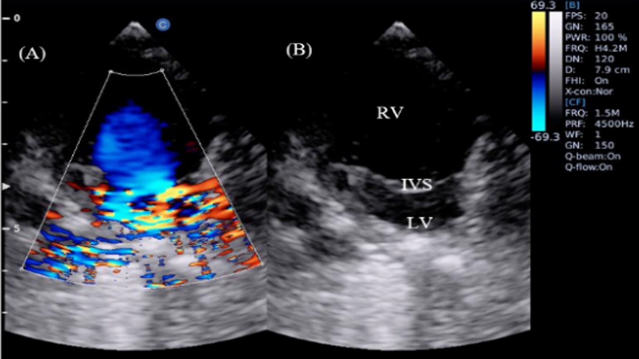
Myxomatous mitral valve disease (MMVD) is a prevalent inherited mitral valve condition. This study aimed to evaluate the clinical manifestation of the cardiac condition MMVD in Poodles. Using ultrasonography (USG), this study examined the characteristics, progression, diagnosis, and treatment of MMVD in an 11-year-old Poodle. This case study was conducted at the Veterinary Teaching Hospital, School of Veterinary Medicine and Biomedical Sciences, utilizing a Chison Ebit60 with an 8–12 MHz curvilinear probe. The dog was positioned in the right parasternal recumbency position, and the long axis (RPLA) and short axis (RPSA) views were obtained. In the evaluation and diagnosis, B-Mode, M-Mode, and Color Flow Doppler (CFD) modes of ultrasonography were performed. The dog presented with coughing, and the physical examination revealed a grade 3–4 murmur. Based on B-mode cardiac monitoring, sinus arrhythmia, mitral valve thickness, and prolapses indicate MMVD. Mitral regurgitation was indicated by a decrease in heart rate and an increase in the left ventricle internal dimension (LVIDd) on the M-mode. Meanwhile, CFD's representation of turbulent flow confirmed mitral regurgitation results. The rise in blood pressure confirmed the presence of hypertension. Class B2 MMVD in Poodles has consequently been diagnosed in this case.
DeFrancesco, T. (2021). POCUS: Heart–Pericardial Effusion and Pericardiocentesis. Point"of"Care Ultrasound Techniques for the Small Animal Practitioner, 417–424.
Fox, P. R. (2012). Pathology of myxomatous mitral valve disease in the dog. Journal of Veterinary Cardiology, 14(1), 103–126.
Hanft, L. M., Fitzsimons, D. P., Hacker, T. A., Moss, R. L., & McDonald, K. S. (2019). The Role of Cardiac MYBPC in Regulating Frank Starling Relationships. Biophysical Journal, 116(3), 116a.
Hezzell, M. (2018). Pathology and prognosis of canine myxomatous mitral valve disease. In Practice, 40(S1), 3–6.
Isayama, N., Uchimura, Y., Sasaki, K., Maeda, E., Takahashi, T., & Watanabe, M. (2022). Reference Values of M-mode Echocardiographic Parameter in Adult Toy Breed Dogs. Frontiers in Veterinary Science, 9.
Kampourakis, T., & Irving, M. (2021). The regulatory light chain mediates inactivation of myosin motors during active shortening of cardiac muscle. Nature Communications, 12(1), 5272.
Keene, B. W., Atkins, C. E., Bonagura, J. D., Fox, P. R., Häggström, J., Fuentes, V. L., Oyama, M. A., Rush, J. E., Stepien, R., & Uechi, M. (2019). ACVIM consensus guidelines for the diagnosis and treatment of myxomatous mitral valve disease in dogs. Journal of Veterinary Internal Medicine, 33(3).
Kim, H. T., Han, S. M., Song, W. J., Kim, B., Choi, M., Yoon, J., & Youn, H. Y. (2017). Retrospective study of degenerative mitral valve disease in small-breed dogs: survival and prognostic variables. Journal of Veterinary Science, 18(3), 369–376.
Laflamme, D. P. (2022). Key nutrients important in the management of canine myxomatous mitral valve disease and heart failure. Journal of the American Veterinary Medical Association, 260(S3), S61–S70.
Noviana, D., & Kurniawan, L. K. L. (2013). Heart size evaluation of Indonesian domestic house cat by motion mode echocardiography imaging. HAYATI Journal of Biosciences, 20(1), 40–46.
Oyama, M. A., Elliott, C., Loughran, K. A., Kossar, A. P., Castillero, E., Levy, R. J., & Ferrari, G. (2020). Comparative pathology of human and canine myxomatous mitral valve degeneration: 5HT and TGF-β mechanisms. Cardiovascular Pathology, 46, 107196.
Öztürk, C., Schueler, R., Weber, M., Welz, A., Werner, N., Nickenig, G., & Hammerstingl, C. (2016). Sympathetic activity in patients with secondary symptomatic mitral regurgitation or end-stage systolic heart failure. Cardiovascular Interventions, 9(19), 2050–2057.
Penninck, D., & d'Anjou, M. A. (1991). Atlas of small animal ultrasonography. John Wiley & Sons.
Summerfield, N. (2018). Simplifying mitral valve disease diagnostics. In Practice, 40, 7–11.
Utami, N. D., & Noviana, D. (2019). Sonogram features of myxomatous mitral valve disease and abdominal organ dissorders in a senior mini pomeranian. ARSHI Veterinary Letters, 3(3), 51–52.
Vezzosi, T., Puccinelli, C., Citi, S., & Tognetti, R. (2021). Two radiographic methods for assessing left atrial enlargement and cardiac remodeling in dogs with myxomatous mitral valve disease. Journal of Veterinary Cardiology, 34, 55–63.
Copyright (c) 2024 Toh Pei Wah, Agus Wijaya, Fitria Senja Murtiningrum, Bintang Nurul Iman, Deni Noviana

This work is licensed under a Creative Commons Attribution-NonCommercial-ShareAlike 4.0 International License.
Authors who publish in this journal agree to the following terms:
1. The journal allows the author to hold the copyright of the article without restrictions;
2. The journal allows the author(s) to retain publishing rights without restrictions;
3. The legal formal aspect of journal publication accessibility refers to Creative Commons Attribution-NonCommercial-ShareAlike 4.0 International License (CC BY-NC-SA).


11.jpg)