Erythrogram Profile of Blood Samples Anticoagulated with Tri-potassium Ethylene Diamine Tetraacetic Acid (K3EDTA) Stored for 48 Hours at 4oC

Downloads
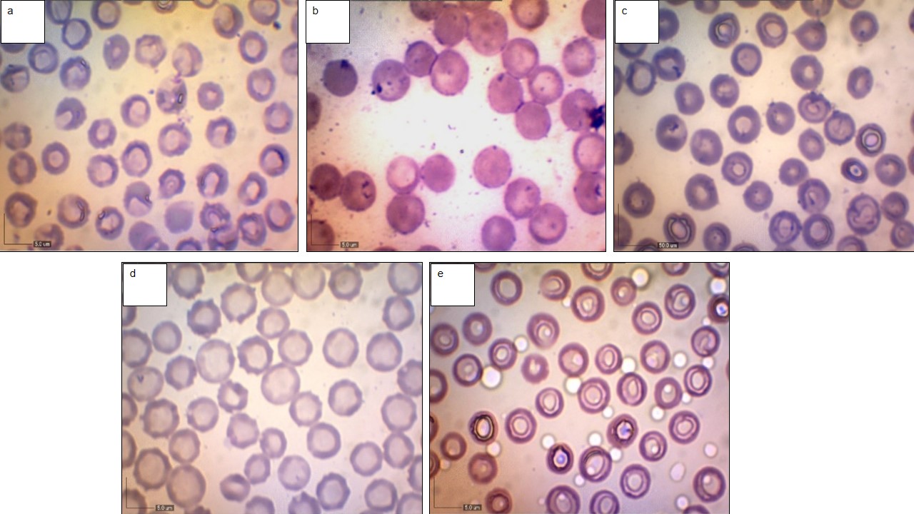
Several pre-analytical variables influence hematological results, including anticoagulant use, storage temperature, and time between blood sample collection and analysis. Delayed sample analysis owing to prolonged storage could result in erythrogram profiles, which could complicate the interpretation of the resulting data. This study investigated the erythrogram profile of tripotassium ethylenediaminetetraacetic acid (K3EDTA) in blood samples stored for 48 h at 4°C. Ten healthy blood samples of Ongole crossbred cattle were collected into K3EDTA tubes from the jugular or coccygeal veins and analyzed for erythrogram profiles (erythrocyte counts, hemoglobin levels, hematocrit value, and erythrocyte morphology). Blood sample analysis for the control (0 h) was performed within ± 1.5 hours after collection, then the samples were refrigerated (4°C) and analyzed at 3, 6, 9, 12, 24, and 48 h. The results showed increased (p < 0.05) erythrocyte counts and hematocrit values after 9–24 and 6–48 h of storage, respectively. There was a significant difference in erythrocyte diameter between 0 h and other time observations (p < 0.05). Echinocytes were observed at 0 h of storage and continued to increase up to 48 h. Hypochromasia was also found at 6 to 48 hours of storage. Therefore, the analysis of blood samples for erythrogram parameters should be performed as soon as possible, preferably within three hours after collection, to ensure clinically reliable results.
Adili, N., Melizi, M., & Belabbas, H. (2016). Species determination using the red blood cells morphometry in domestic animals. Veterinary World, 9(9), 960–963.
Alagbe, E. E., Susu, A. A., & Dosunmo, A. O. (2013). Sickle cell disease (scd) management: a theoretical review. IJRRAS, 16(3), 483–500.
Alan, W. H. B. (2006). Tietz Clinical Guide to Laboratory Test. St. Louis Missouri: Saunders Elsevier.
Ali, N. T. (2017). Effect of storage at temperature (4°C) on complete blood count parameters. Journal of Cancer Treatment and Research, 5(2), 7–10.
Andriyani, Y., Kusumaningrum, S. B. C., & Sepvianti, W. (2019). Gambaran jumlah eritrositpada whole blood selama 30 hari penyimpanan di PMI Kabupaten Sleman, Yogyakarta. Conference on Research and Community Services, 1(1), 463–467.
Andriyanto, Rahmadani, Y. S., Satyaningsih, A. S., & Abadi, S. (2010). Gambaran hematologi domba selama transportasi: peran multivitamin dan meniran. Jurnal Ilmu Peternakan Indonesia, 15(3), 134–136.
Antwi-Baffour, S., Quao, E., Kyeremeh, R., & Mahmood, S. A. (2013). Prolong storage of blood in EDTA has an effect on the morphology and osmotic fragility of erythrocytes. International Journal of Biomedical Science and Engineering, 1(2), 20–23.
Bain, B. J. (2014). Hematologi: Kurikulum Inti. Edisi ke-20. Jakarta: EGC.
Bijanti, R., Yuliani, M. G. D., Wahjuni, R. S., & Utomo, R. B. (2010). Buku Ajar Patologi Klinik Veteriner. Edisi ke-1. Surabaya: Universitas Airlangga Press.
Brigden, M. L., & Dalal, B. I. (1999). Cell counter-related abnormalities. Laboratory Medicine, 30(5), 325–334.
Britton, C. J. C. (1963). Disorders of blood. 9th edition. London: J and A Churchill Ltd.
Brockus, C. W. (2011). Erythrocytes. In Latimer, K. S. (Ed) Duncan and Prasse's Veterinary Laboratory Medicine: Clinical Pathology. 5th edition. Chichester: Wiley.
Christopher, M. M., Hawkins, M. G., & Burton, A. G. (2014). Poikilocytosis in rabbits: prevalence, type, and association with disease. PLOS ONE, 9(11), 1–11.
Cunningham, J. G. (2002). Texbook of Veterinary Physiology. St. Louis Missouri: Saunders Company.
Coles, E. H. (1986). Veterinary Clinical Pathology. 4th edition. Philadelphia: WB Saunders Company.
Cora, M. C., King, D., Betz, L. J., Wilson, R., & Travlos, G. S. (2012). Artifactual changes in sprague– dawley rat hematologic parameters after storage of samples at 3°C and 21°C. Journal of the American Association for Laboratory Animal Science, 51(5), 616–621.
Corwin, E. J. (2009). Hematologi Klinik Ringkas. Jakarta: EGC.
Daves, M., Zagler, E. M., Cemin, R., Gnech, F., Joos, A., Platzgummer, S., & Lippi, G. (2015). Sample stability for complete blood cell count using the Sysmex XN hematological analyser. Blood Transfus, 13(4), 576–82.
Dewi, A. K. S, Mahardika, I. G., & Dharmawan, N. S. (2018). Toral eritrosit, kadar hemoglobin, nilai hematokrit sapi bali lepas sapih diberi pakan kandungan protein dan energi berbeda. Indonesia Medicus Veterinus. 7(4):413–421.
Ekanem, A. P., Udoh, A. J., & Inyang-Etoh, A. P. (2012). Effect of different anticoagulants on hematological parameters of oreochromis niloticus. IJSAT, 2(6), 17–20.
Fitria, L., Illiy, L. L., & Dewi, I. R. (2016). Pengaruh antikoagulan dan waktu penyimpanan terhadap profil hematologis tikus (Rattus norvegicus Berkenhout, 1769) galur wistar. Biosfera, 33(1), 22–30.
Frandsond, R. D. (1993). Anatomi dan Fisiologi Ternak. Edisi ke-4. Yogyakarta: UGM Press.
Gandasoebrata, R. (2013). Penenutun Laboratorium Klinik. Jakarta: Dian Rakyat.
Guyton, A. C. (1991). Fisiologi Kedokteran. Edisi ke-5. Dharma, A., penerjemah. Jakarta: Penerbit Buku Kedokteran EGC.
Guyton, A. C., & Hall, J. E. (2008). Buku Ajar Fisiologi Kedokteran. Edisi ke-11. Irawati, Ramadani, D., & Indriyani, F., penerjemah. Jakarta: Penerbit Buku Kedokteran EGC.
Hartina, H., Garini, A., & Tarmizi, M. I. (2018). Perbandingan teknik homogenisasi darah EDTA dengan teknik inversi dan teknik angka delapan terhadap jumlah trombosit. Jurnal Kesehatan Poltekes Palembang, 13(2), 150–153.
Harvey, J. W. (2012). Veterinary Hematology: A Diagnostic Guide and Color Atlas. St. Louis Missouri: Elsevier Saunders.
Hidayatulloh, K. N. (2021). Komparasi hasil jumlah eritrosit pada volume 1 CC 2 CC dan 3 CC darah tabung K3EDTA setelah 2 jam penyimpanan suhu ruang AC (air conditioner) 18–22oC. [Karya tulis ilmiah]. Jurusan Analis Kesehatan. Politeknik Kesehatan Kementerian Kesehatan.
Hutomo, F. P. (2011). Dasar Dasar Transfusi Darah. Jakarta: WIMI.
Keohane, E. M., Smith, L. J., & Walenga, J. M. (2015). Rodaks's Hematology: Clinical Principles and Applications. 5th edition. St. Louis Missouri: Elsevier Saunders.
Kiswari, R. (2014). Hematologi dan Transfusi. Jakarta: Erlangga.
Kosasih, E. N., & Kosasih, A. S. (2008). Tafsiran Hasil pemeriksaan Laboratorium Klinik. Edisi ke-2. Tangerang: Karisma Publishing Group.
Kristanto, D., & Septiyani. (2023). Comparison of Hematological Levels of Simmental-Ongole Crossbreed (SimPO) and Ongole Crossbreed (PO) Cattle Reared Semi-Intensively. Jurnal Medik Veteriner, 6(2), 237–243.
Li, X., Feng, H., Wang, Y., Zhou, C., Jiang, W., Zhong, M., & Zhou, J. (2018). Capture of red blood cells onto optical sensor for rapid ABO blood group 81 typing and erythrocyte counting. Sensors and Actuators B Chemical, 262, 411–417.
Lindstrom, N. M., Moore, D. M., Zimmerman, K., & Smith, S. A. (2015). Hematologic assessment, and gerbils: blood sample collection and blood cell identification. Clinics in Laboratory Medicine, 35(3), 629–640.
Majid, R. A., Septiyani, Gradia, R., Rosdianto, A. M., & Hidayatik, N. (2023). Hematological Profile in Dairy Cattle with Foot and Mouth Diseases in Lembang, West Bandung. Jurnal Medik Veteriner, 6(3), 381–389.
Megarani, D. V., Hardian, A. B., Arifianto, D., Santosa, C. M., & Salasia, S. I. O. (2020). Comparative morphology and morphometry of blood cells in zebrafish (Danio rerio), common carp (Cyprinus carpio carpio), and tilapia (Oreochromis niloticus). Journal of the American Association for Laboratory Animal Science, 59(6), 1–8.
Mindray. (2006). BC-3000 Auto Hematology Analyzer. Shenzhen: Mindray Building.
Mingoas, J. P. K., Somnjom, D. E., Moffo, F., Mfopit, Y. M., & Awah-Ndukum, J. (2020). Effect of duration and storage temperature on hematological and biochemical parameters of Clinically Healthy Gudali Zebu in Ngaoundere, Cameroon. Singapore Journal of Scientific Research, 10(2), 190–196.
Mulyadi, A., Triya, M. L., Barradillah, A., Nuzul, A., Muttaqien, & Fakhrurrazi. (2015). Jumlah eritrosit dan nilai hematokrit sapi aceh dan sapi Bali di Kecamatan Leumbah Seulawah Kabupaten Aceh Besar. Jurnal Medika Veterinaria, 9(2), 15–118.
Nair, M., & Peate, I. (2013). Fundamentals Of Applied Pathophysiology: An Essential Guide Nursing And Healthcare Students. 2th edition. Chichester: John Wiley and Sons Limited.
Novel, S., Apriyani, R. K., Setiadi, H., & Safitri, R. (2012). Biomedik. Jakarta: Trans Info Media.
Ozmen, S. U., & Ozarda, Y. (2021). Stability of hematological analytes during 48 hours storage at three temperatures using cell-dyn hematology analyzer. Journal of Medical Biochemistry, 40(3), 252–260.
Price, S. A., & Wilson, L. M. (2006). Patophysiology Clinical Conceps of Disease Processes. Edisi ke-4. Jakarta: Penerbit Buku Kedokteran EGC.
Purnama, M. T. E., Hendrawan, D., Wicaksono, A. P., Fikri, F., Purnomo, A., & Chhetri, S. (2021). Risk factors, hematological and biochemical profile associated with colic in Delman horses in Gresik, Indonesia. F1000Research, 10, 950.
Purnama, M. T. E., Dewi, W. K., Prayoga, S. F., Triana, N. M., Aji, B. S. P., Fikri, F., & Hamid, I. S. (2019). Preslaughter stress in banyuwangi cattle during transport. Indian Veterinary Journal, 96(12), 50-52.
Putri, U. A., Adang, D., Betty, N., & Ganjar, N. (2019). Waktu simpan darah antikoagulan K2EDTA dan K3EDTA terhadap parameter eritrosit. Jurnal Riset Kesehatan, 11(2), 175–189.
Rahmanitarini, A., Hernaningsih, Y., & Indrasari, Y. N. (2019). The stability of sample storage for complete blood count (CBC) toward the blood cell morphology. Bali Medical Journal, 8(2), 391–395.
Riswanto. (2013). Pemeriksaan Laboratorium Hematologi. Yogyakarta: Alfamedia.
Saputro, A. A., & Lestari, C. R. (2021). Pengaruh waktu penyimpanan darah terhadap kadar hemoglobin pada komponen whole blood darah donor. Jurnal Analis Laboratorium Medik, 6(2), 50–56.
Schapkaitz, E., & Pillay, D. (2015). Prolonged storage-induced changes in hematology parameters referred for testing. African Journal of Laboratory Medicine, 4(1), 208–215.
Schalm, O. W., Carrol, E. J., & Join, N. C. (1975). Phisiology properties of celular and chemical constituens of blood. Didalam: Swenson, M. J. (Ed) Dukes Physiology of Domestic Animals. Ithaca: Cornell University Press.
Septiarini, A. A. I. A., Suwiti, N. K., & Suartini, I. G. A. A. (2020). Nilai hematologi total eritrosit dan kadar hemoglobin sapi bali dengan pakan hijauan organik. Buletin Veteriner Udayana, 12(2), 144–149.
Septiyani, Majid, R. A., Gradia, R., Setiawan, I., Yantini, P., & Novianti, A. N. (2023). Peripheral Blood Smear Analysis for Cattle with Foot and Mouth Diseases in Lembang, West Bandung. Jurnal Medik Veteriner, 6(3), 319–325.
Simeonov, R. (2012). Nuclear morphometry in cytological specimens of canine ceruminous adenomas and carcinomas. Veterinary and Comparative Oncology, 10(4), 246–251.
Smith, J. B., & Mangkoewidjojo, S. (1988). Pemeliharaan Pembiakan dan Penggunaan Hewan Percobaan di daerah Tropis. Jakarta: Universitas Indonesia Press.
Stokol, T., Priest, H., Behling-Kelly, E., & Babcock, G. (2014). Samples for Hematology. Animal Health Diagnostic Center. Clinical Pathology Laboratory. College of Veterinary Medicine, Cornell University. Ithaca, New York.
Voigt, G. L., & Swist, S. L. (2011). Hematology Techniques And Concepts For Veterinary Technicians. 2th edition. Chichester: WileyBlackwell.
Wismaya, H. S. (2019). Pengaruh morfologi dan jumlah sel darah merah terhadap karakteristik nilai impedansi whole blood cell menggunakan metode spektroskopi impedansi listrik. [Tesis]. Malang: Universitas Brawijaya.
Wood, D., & Quiroz-Rocha, G. F. (2010). Normal hematology of cattle. Didalam: Weiss, D., Wardrop, K. J., (Ed) Schalm's Veterinary Hematology. Edisi ke-6. Philadelphia: Wiley-Blackwell.
Copyright (c) 2024 Putri Azzahrah, Anita Esfandiari, Arief Purwo Mihardi, Sus Derthi Widhyari, Retno Wulansari, Putri Indah Ningtias

This work is licensed under a Creative Commons Attribution-NonCommercial-ShareAlike 4.0 International License.
Authors who publish in this journal agree to the following terms:
1. The journal allows the author to hold the copyright of the article without restrictions;
2. The journal allows the author(s) to retain publishing rights without restrictions;
3. The legal formal aspect of journal publication accessibility refers to Creative Commons Attribution-NonCommercial-ShareAlike 4.0 International License (CC BY-NC-SA).


11.jpg)


