Surgical Management of Biliary Duct Hamartoma in a Cat: A Case Report

Downloads
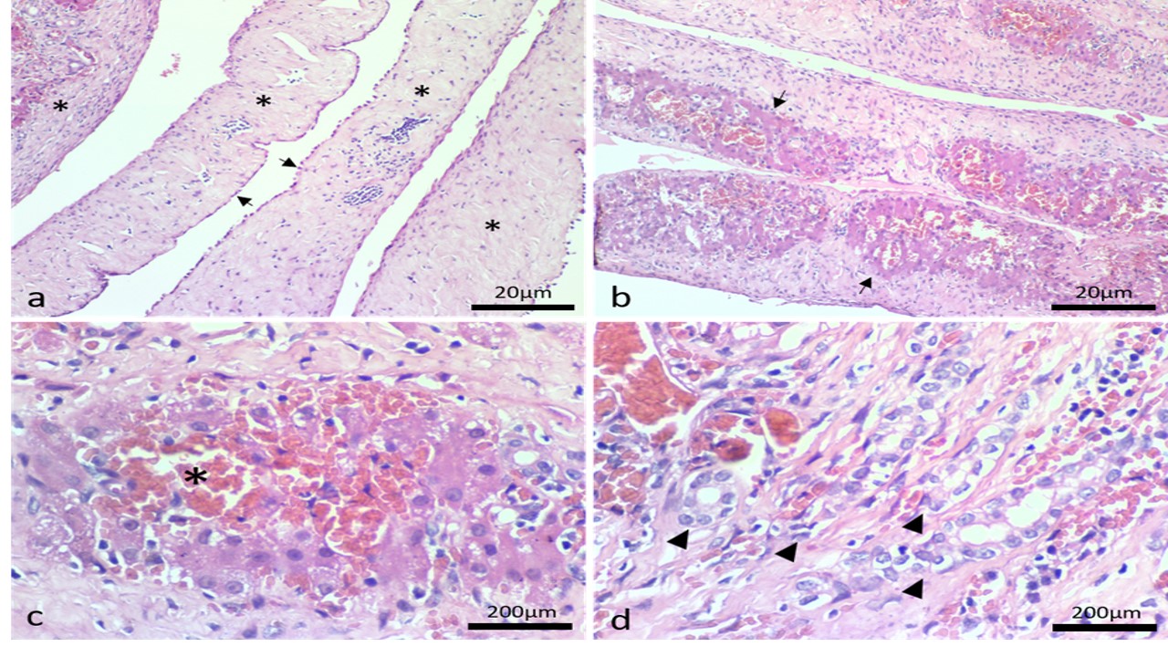
Cysts in the liver or biliary duct are uncommon in veterinary medicine. A multiloculated, fluid-filled liver cyst measuring 18 cm in diameter was detected in a two-year-old spayed female mixed-breed cat via radiography and computed tomography. The cyst was attached to the medial lobe aspect of the liver and continued with the gall bladder. Cystectomy and omentopexy were performed, and the resected cyst was examined histologically. Histologic analysis revealed variable-sized cystic spaces lined by low simple cuboidal and attenuated epithelium. The cyst wall was composed of thick collagenous stroma containing entrapped islands of the hepatic parenchyma, which included atrophied hepatocytes, dilated sinusoidal spaces filled with erythrocytes, and randomly distributed hyperplastic bile ducts. These histologic findings were consistent with biliary duct hamartoma. The cat had an uneventful recovery, and no recurrence was observed one-year post-surgery.
Andrade, R. L., Dantas, A. F., Pimentel, L. A., Galiza, G. J., Carvalho, F. K., Costa, V. M., & Riet-Correa, F. (2012). Platynosomum fastosum-induced cholangiocarcinomas in cats. Veterinary Parasitology, 190(1–2), 277–280.
Bonelli, P., Masu, G., Dei Giudici, S., Pintus, D., Peruzzu, A., Piseddu, T., Santucciu, C., Cossu, A., Demurtas, N., & Masala, G. (2018). Cystic echinococcosis in a domestic cat (Felis catus) in Italy. Échinococcose kystique chez un chat domestique (Felis catus) en Italie. Parasite (Paris, France), 25, 25.
Center, S. A. (2023). ‘Miscellaneous Disorders of the Bile Ducts in Small Animals’, in MSD Veterinary Manual. Available: https://www.msdvetmanual.com. (Accessed: 30th June 2024)
De Bosschere, H., & Ducatelle, R. (1999). Bile duct hamartoma in a calf. The Veterinary Record, 144(8), 210–211.
Kim, K., Kim, H., Eom, K., & Kim, H. (2021). Surgical management and long-term follow-up of a giant hepatic cyst with an internal septum in a cat. Journal of Veterinary Clinics, 38(6), 295–298.
Mao, D., Song, X., Ma, D., Hu, S., Zhang, Z., Wang, J., & He, X. (2023). Bile duct hamartoma in a dog. Journal of Comparative Pathology, 207, 45–49.
Moon, S. J., Kim, J. W., Sur, J. H., Jeong, S. W., & Park, H. M. (2011). Biliary cystadenoma in a Maltese dog: clinical and diagnostic findings. The Journal of Veterinary Medical Science, 73(12), 1677–1679.
Naga, M. A., Abd El Aal, A. A., Mousa, A. S., & Saber, H. S., (2020). Omentopexy in sleeve gastrectomy and its effect on postoperative complications. The Egyptian Journal of Surgery, 39(4), 1208.
Naghi, R., Bertran, J., Spoldi, E., Dark, M. J., de Oliveira, H. H., Souza, C., & Maxwell, E. A. (2023). Multiple biliary duct hamartomas in a cat resulting in a hepatic mass: A case report. Veterinary Medicine and Science, 9(4), 1441–1445.
Radlinsky, M. A., & Fossum, T. W. (2019). Surgery of the liver. In Fossum, T. W. (Ed) Small Animal Surgery. 5th edition. Philadelphia PA: Elsevier. pp: 540–570.
Roberts, M. L., Rine, S., & Lam, A. (2018). Caroli's-type ductal plate malformation and a portosystemic shunt in a 4-month-old kitten. JFMS Open Reports, 4(2), 2055116918812329.
Schreeg, M. E., Miller, S. A., & Cullen, J. M. (2021). Choledochal cyst with secondary cholangitis, choledochitis, duodenal papillitis, and pancreatitis in a young domestic shorthair cat. Journal of Veterinary Diagnostic Investigation, 33(4), 782–787.
Sheikh, A. A. E., Nguyen, A. P., Leyba, K., Javed, N., Shah, S., Deradke, A., Cormier, C., Shekhar, R., & Sheikh, A. B. (2022). Biliary Duct Hamartomas: A Systematic Review. Cureus, 14(5), e25361.
Starost M. F. (2007). Solitary biliary hamartoma with cholelithiasis in a domestic rabbit (Oryctolagus cuniculus). Veterinary Pathology, 44(1), 92–95.
Venkatanarasimha, N., Thomas, R., Armstrong, E. M., Shirley, J. F., Fox, B. M., & Jackson, S. A. (2011). Imaging features of ductal plate malformations in adults. Clinical Radiology, 66(11), 1086–1093.
Copyright (c) 2025 Nur Ainina Ab Manap, Tey Yu Chong, Erni Wati Mohd Arip, Rozanaliza Radzi, Muhamad Alif Zakaria, Nur Diyana Mohamad Tahir, Nurul Izzati Uda Zahli

This work is licensed under a Creative Commons Attribution-NonCommercial-ShareAlike 4.0 International License.
Authors who publish in this journal agree to the following terms:
1. The journal allows the author to hold the copyright of the article without restrictions;
2. The journal allows the author(s) to retain publishing rights without restrictions;
3. The legal formal aspect of journal publication accessibility refers to Creative Commons Attribution-NonCommercial-ShareAlike 4.0 International License (CC BY-NC-SA).


11.jpg)