Comparative Scanning Electron Microscopy Study on Scale Variations in Indonesian Cultivated Koi Fish (Cyprinus rubrofuscus Lacepede, 1803)

Downloads
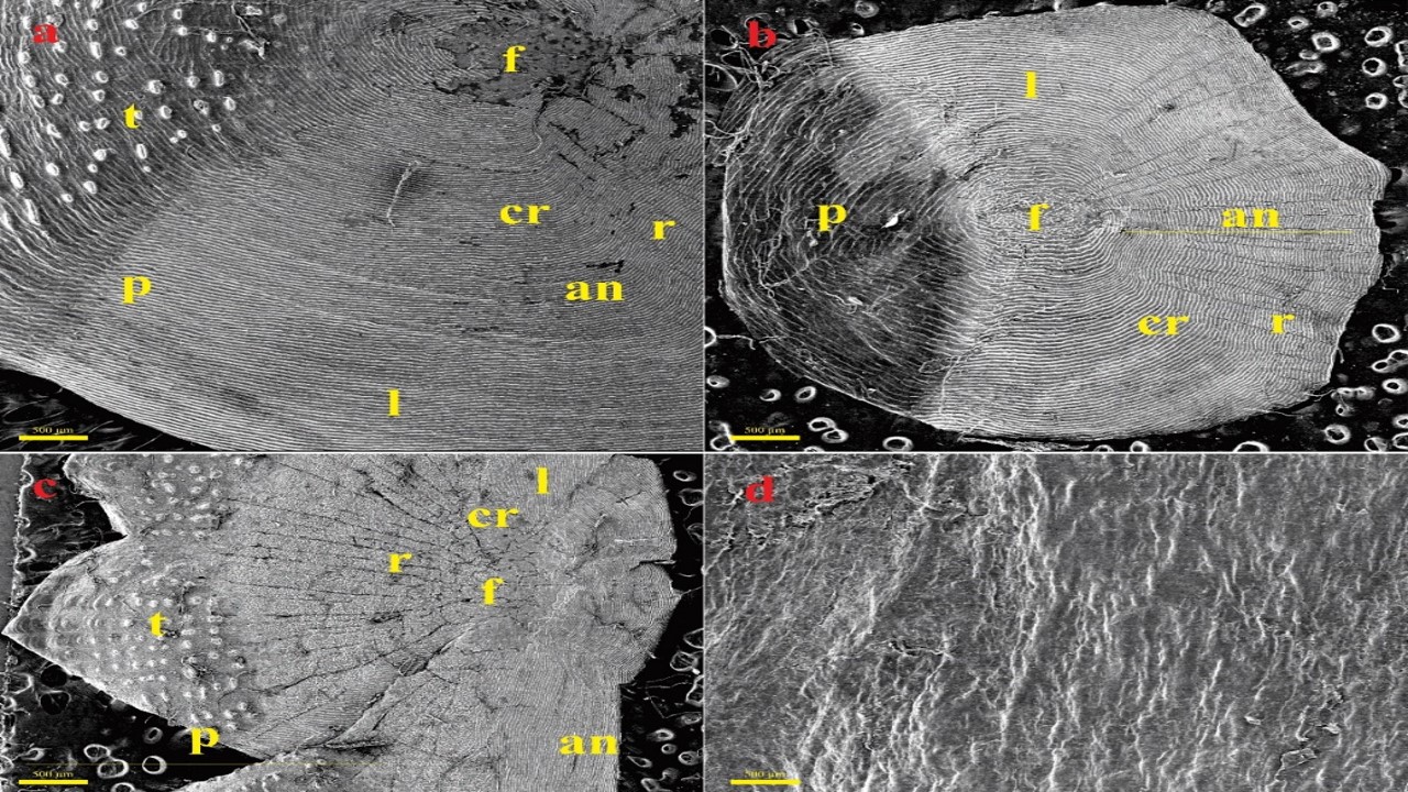
Koi (Cyprinus rubrofuscus Lacepede, 1803) is a highly favored ornamental fish due to its beauty and wide range of variations in Indonesia, categorized by color, patterns, and scales. Some variants are distinguished by color, while others, such as Ginrin, Doitsu, and Shusui, have unique scale types. Despite visible differences, microscopic scale variations remain unexplored. SEM studies in other fish species offer insights into scale ultrastructure, providing opportunities for comparison. Therefore, this study aimed to uncover the microscopic structure of four types of koi fish, namely Doitsu, Ginrin, Shusui, and common scale. Koi fish were obtained from breeders in Yogyakarta, Indonesia, and acclimatized in an aquarium. Scale extraction was conducted under anesthesia using MS-222 and the cleaned scales were then subjected to dehydration, fixation, and affixed to double-sided adhesive tape for SEM analysis. Furthermore, coating with conductive gold enabled observation using SEM at 10 kV, allowing examination of scale features such as focus, radii, circuli, and lepidonts at various magnifications. The results showed that based on SEM analysis, significant differences were observed in scale structures among koi variants. At low magnification, differences in tubercles and lepidonts were observed, particularly between common and Ginrin scale types. Shusui scale showed unique characteristics with a closer arrangement of circuli and distinctive lepidont shapes. At higher magnification, clearer details of radii, circuli, and lepidonts were observed, further highlighting the differences among koi variants. SEM provides crucial insights into the morphology of scales in koi fish variants, showing unseen macroscopic differences and distinct features such as tubercles and lepidont frequency.
Al Jufaili, S. M., Masoumi, A. H., Esmaeili, H. R., Jawad, L. A., & Teimori, A. (2021). Morphological and microstructural characteristics of scales in longnose goby Awaous jayakari (Teleostei: Gobiidae): Light and scanning electron microscopy approaches. Microscopy Research and Technique, 84(12), 3128–3149.
Al Jufaili, S. M., Echreshavi, S., & Esmaeili, H. R. (2023). Scales surface topography: Comparative ultrastructural and decorative characteristics of a modern elasmoid fish scales in a cyprinid fish, Garra shamal (Teleostei: Cyprinidae) using digital optical light and scanning electron microscope imaging. Microscopy Research and Technique, 86(1), 97–114.
Andrian, K. N., ’Aisy, N. R., Novindasari, B. B. M., Nurrahmi, I. A., Santi, M. D., & Haryanto, A. (2023). Random Amplified Polymorphic DNA-Polymerase Chain Reaction Analysis of Four Koi Fish (Cyprinus carpio var. koi) Variants from Yogyakarta, Indonesia. IOP Conference Series: Earth and Environmental Science, 1174, 1–7.
Andrian, K. N., Wihadmadyatami, H., Wijayanti, N., Karnati, S., & Haryanto, A. (2024). A comprehensive review of current practices, challenges, and future perspectives in Koi fish (Cyprinus carpio var. koi) cultivation. Veterinary World, 17(8), 1846–1854.
Ansari, S., Chavan, S., & Shaikh, Y. (2021). Scanning electron microscopy of scales in two edible fishes Notopterus kapirat and Etroplus suratensis for its application in taxonomy. International Journal of Fisheries and Aquatic Studies, 9(4), 315–319.
Aysi, N. R., Santi, M. D., Andrian, K. N., & Haryanto, A. (2022). Molecular fish sexing on Kohaku Koi (Cyprinus carpio) based on ArS.9–15 gene amplification by PCR method. IOP Conference Series: Earth and Environmental Science, 976(1), 1–6.
Clark, A. J., & Uyeno, T. A. (2024). The biomechanics of fish skin. In Encyclopedia of Fish Physiology. Elsevier, pp: 476–498.
de Kock, S., & Gomelsky, B. (2015). Japanese Ornamental Koi Carp: Origin, Variation and Genetics. In Biology and Ecology of Carp. CRC Press, pp: 27–53.
Dey, S., Biswas, S. P., & Dey, S. (2015). Scanning electron microscopy of scale microstructures in Channa barca, a poorly known snake head fish in regard to taxonomy. Microscopy, 64(5), 351–359.
Dey, S., Biswas, S. P., Dey, S., & Bhattacharyya, S. P. (2014). Scanning electron microscopy of scales and its taxonomic application in the fish genus Channa. Microscopy and Microanalysis, 20(4), 1188–1197.
Dhamayanti, Y., Khairunnisa, H. K., Zahrudin, E., Bayram, M., & Suciyono, S. (2024). Immune responses of club cells in fish: A review. Jurnal Medik Veteriner, 7(2), 407–412.
Ding, Y., Zou, M., & Guo, B. (2024). Genomic signatures associated with recurrent scale loss in cyprinid fish. Integrative Zoology, 00, 1–16.
Echreshavi, S., Esmaeili, H. R., Teimori, A., Safaie, M., & Owfi, F. (2021). Hidden morphological and structural characteristics in scales of mullid species (Teleostei: Mullidae) using light and scanning electron digital imaging. Microscopy Research and Technique, 84(11), 2749–2773.
Esmaeili, H. R., & Gholami, A. (2011). Scanning Electron Microscopy of the scale morphology in Cyprinid fish, Rutilus frisii kutum Kamenskii, 1901 (Actinopterygii: Cyprinidae). Iranian Journal of Fisheries Sciences, 10(1), 155–166.
Faal, S. A., Esmaeili, H. R., & Teimori, A. (2024). Scale Characteristics of Six Fish Species of the Genus Cyprinion (Teleostei: Cypriniformes): A Microscopic Analysis. Microscopy and Microanalysis, 30(4), 771–792.
Fikri, F., Wardhana, D. K., Purnomo, A., Khairani, S., Chhetri, S., & Purnama, M. T. E. (2022). Aerolysin gene characterization and antimicrobial resistance profile of Aeromonas hydrophila isolated from milkfish (Chanos chanos) in Gresik, Indonesia. Veterinary World, 15(7), 1759–1764.
Ghods, S., Waddell, S., Weller, E., Renteria, C., Jiang, H. Y., Janak, J. M., Mao, S. S., Linley, T. J., & Arola, D. (2020). On the regeneration of fish scales: structure and mechanical behavior. Journal of Experimental Biology, 223(10), jeb211144.
Gomelsky, B., Delomas, T. A., Schneider, K. J., Anil, A., & Warner, J. L. (2015). Inheritance of Sparkling Scales (Ginrin) Trait in Ornamental Koi Carp. North American Journal of Aquaculture, 77(3), 312–317.
Gu, H., Wang, H., Zhu, S., Yuan, D., Dai, X., & Wang, Z. (2023). Interspecific differences and ecological correlations between scale number and skin structure in freshwater fishes. Current Zoology, 69(4), 491–500.
Murcia, S., Lavoie, E., Linley, T., Devaraj, A., Ossa, E. A., & Arola, D. (2017). The natural armors of fish: A comparison of the lamination pattern and structure of scales. Journal of the Mechanical Behavior of Biomedical Materials, 73, 17–27.
Novindasari, B. M., Nurrahmi, I. A., & Andrian, K. N. (2024). Molecular fish sexing on Taisho Sanshoku Koi (Cyprinus carpio) based on ArS.9-15 gene amplification using PCR method. Jurnal Medik Veteriner, 7(2), 255–263.
Raffealla, N., & Bhuyan, R. (2020). Comparative study of fish scale using scanning electron microscopy in two Cyprinid fishes (Neolissochilus hexagonolepis and Neolissochilus hexastichus) found in Meghalaya, North-East India. International Journal of Life Sciences, 8(1), 77–82.
Scott, E., Edgley, D. E., Smith, A., Joyce, D. A., Genner, M. J., Ioannou, C. C., & Hauert, S. (2023). Lateral line morphology, sensory perception and collective behaviour in African cichlid fish. Royal Society Open Science, 10(1), 221478.
Suciyono, S., Mukti, A. T., Kenconojati, H., Ulkhaq, M. F., Fasya, A. H., Lamadi, A., Imlani, A., & Rao Mariah, S. (2023). Color brightness and growth levels of goldfish (Carassius auratus) reared with different light spectrums. Jurnal Medik Veteriner, 6(2), 250–255.
Ujjania, N. C., & Jaiswar, A. K. (2024). Scale morphology an additional tool for taxonomy and fish identification with reference to Nemipteridae fishes (N. japonicus, N. bipunctatus and N. randalli). Flora and Fauna Jhansi, 30(2), 11–18.
Vernerey, F. J., & Barthelat, F. (2010). On the mechanics of fishscale structures. International Journal of Solids and Structures, 47(17), 2268–2275.
Copyright (c) 2025 Krisna Noli Andrian, Hevi Wihadmadyatami, Nastiti Wijayanti, Srikanth Karnati, Aris Haryanto

This work is licensed under a Creative Commons Attribution-NonCommercial-ShareAlike 4.0 International License.
Authors who publish in this journal agree to the following terms:
1. The journal allows the author to hold the copyright of the article without restrictions;
2. The journal allows the author(s) to retain publishing rights without restrictions;
3. The legal formal aspect of journal publication accessibility refers to Creative Commons Attribution-NonCommercial-ShareAlike 4.0 International License (CC BY-NC-SA).


11.jpg)