Study of Laboratory Diagnosis of Colibasilosis Infection In Local Hen In Surabaya

Downloads


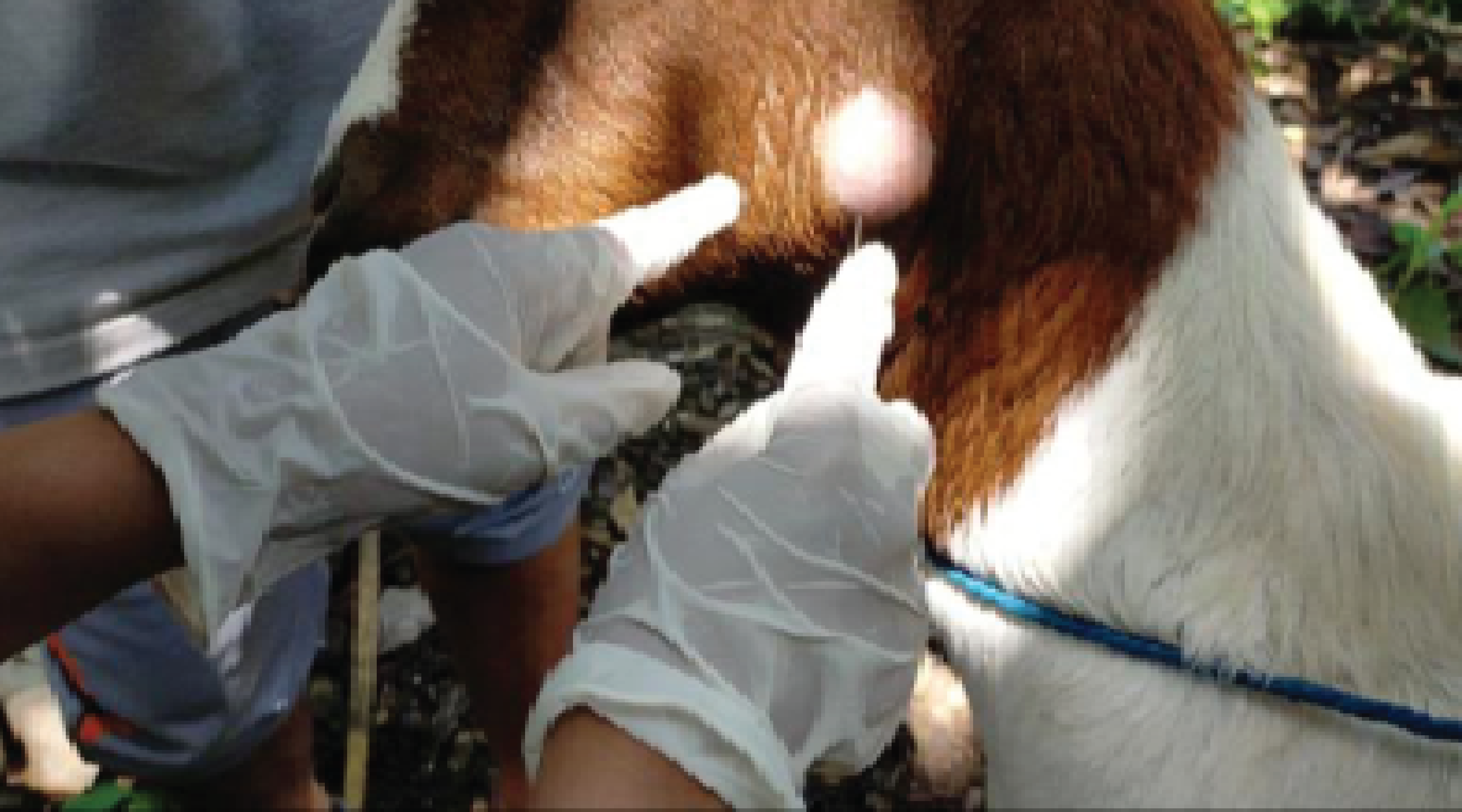
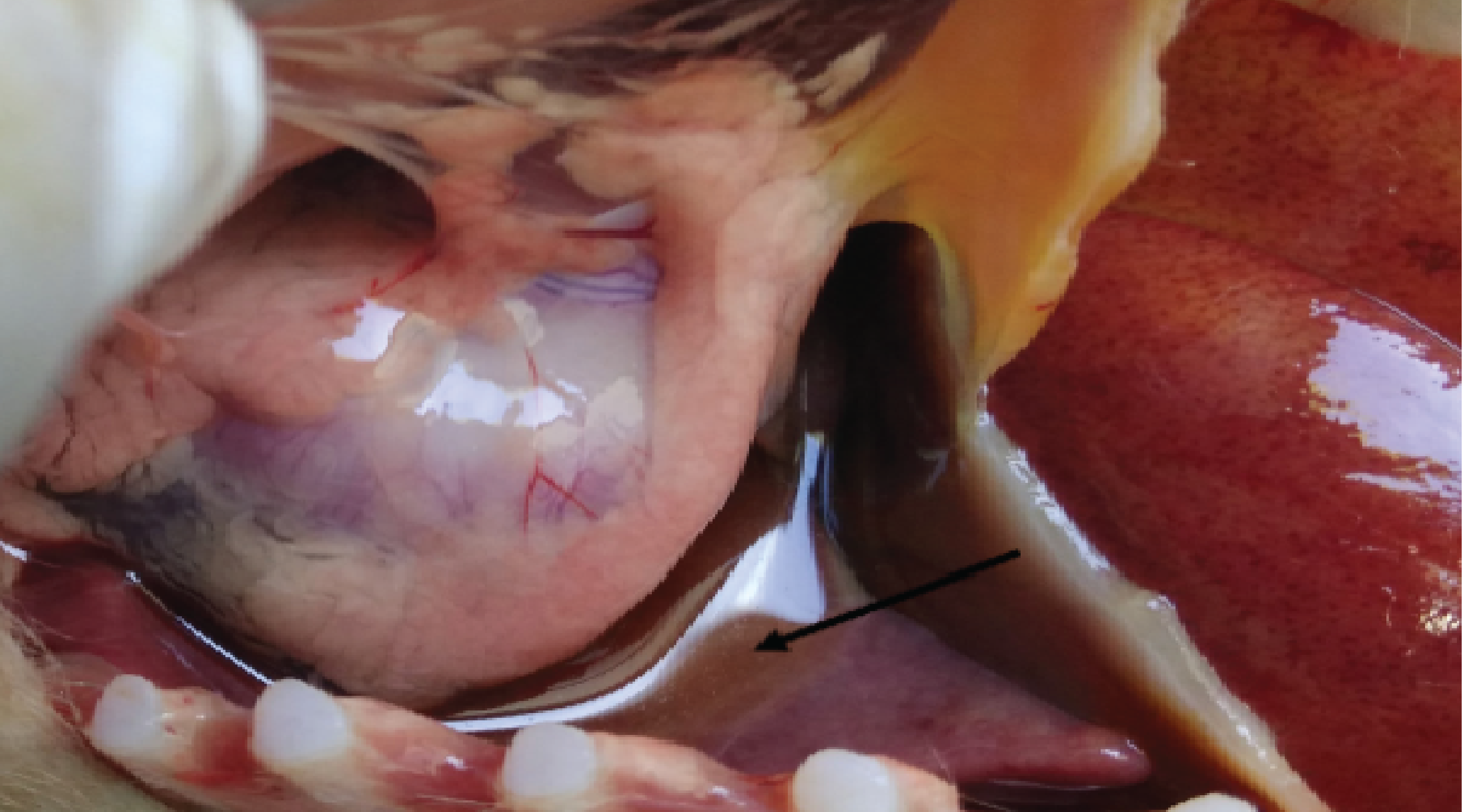
Background: On November 18, 2022, euthanasia and hen necropsy were conducted (protocol number A-164) in the veterinary pathology laboratory, Faculty Of Veterinary Medicine, Wijaya Kusuma University Surabaya. The hen belongs to a local farmer located at Sawahan, Surabaya. Based on the anamnesis, it is known that the hen was four months old and weighed 1.4 kg. The population consists of eight birds, and one of them was sick, never treated or vaccinated, in a cage made of bamboo, corn fodder, rice bran, and drinking water from tap water. Clinical symptoms show a decreased appetite for the last day, watery eyes, runny nose, lethargy, and diarrhea with feces green mixed with white colors. The initial diagnosis of the chicken was infected with Salmonella sp. with a differential diagnosis of Escherichia coli. Purpose: To diagnose bacterial disease in local chickens with a pathological examination, parasitological examination, microbiological isolation, and clinical pathology for hematology. Result: The results of the anatomic pathology examination showed that the chickens had hemorrhagic tracheitis, hemorrhagic necrotic pneumonia, hemorrhagic necrotic enteritis, hemorrhagic necrotic hepatitis, and hemorrhage in the proventriculus. Parasite identification was found in Raillietina sp. and Ornithonyssus sylviarum. Isolation of bacteria was found in Escherichia coli from the duodenum organ. The chickens had hypoproteinemia, poikilocytosis teardrop and rouleaux, leukocytosis with heterophilia, and monocytosis. Conclusion: Based on the examination associated with the results of anamnesis and examination of clinical symptoms, the chickens were diagnosed with Escherichia coli infection together with Raillietina sp. and Ornithonyssus sylviarum infestations.
Aini, M., Rahayuni, S., Mardina, V., Quranayati, Q., and Asiah, N., 2021. Bakteri Lactobacillus spp dan Peranannya Bagi Kehidupan. Jurnal Jeumpa, 8 (2), 614–624.
Andiarsa, D., Hidayat, S., Setyaningtyas, D.E., and Sudarmawan, S., 2014. Gambaran Kerusakan Mukosa Usus Mencit (Mus musculus) pada Infeksi Escherichia coli. Jurnal Vektor Penyakit, 8 (2), 53–60.
Astuti, N., 2012. Kinerja Ayam Kampung Dengan Ransum Berbasis Konsentrat Broiler. Jurnal AgriSains, 4 (5), 51–58.
Bambang, A.G., Fatimawali, F., and Kojong, N.S., 2014. Analisa Cemaran Bakteri Coliform dan Identifikasi E.coli pada Air Isi Ulang dari Depot di Kota Manado. Pharmacon, 3 (3), 325–334.
Baratawidjaja, K.G. and Rengganis, I., 2012. Imunologi Dasar. 10th ed. Jakarta: Fakultas Kedokteran Universitas Indonesia.
Darsana, I.G.O., Besung, I.N.K., and Mahatmi, H., 2012. Potensi Daun Binahong (Anredera Cordifolia (Tenore) Steenis) dalam Menghambat Pertumbuhan Bakteri Escherichia Coli secara In Vitro. Indonesia Medicus Veterinus, 1 (3), 337–351.
Daud, M., Yaman, M.A., and Zulfan, Z., 2019. Gambaran Histopatologi dan Populasi Bakteri Asam Laktat pada Duodenum Ayam Pedaging yang Diberi Sinbiotik dan Diinfeksi Escherichia coli. Jurnal Veteriner, 20 (3), 307-315
Fahreza, R.A. and Sugiharto, S., 2020. Perbandingan Total Leukosit dan Leukosit Diferensial Ayam Broiler Pada Dataran Tinggi dan Rendah. Journal of Animal Research Aoolied Sciences, 2 (1), 22–28.
Fatiqin, A., Novita, R., and Apriani, I., 2019. Pengujian Salmonella dengan Menggunakan Media SSA dan E.coli Menggunakan Media EMBA pada Bahan Pangan. Jurnal Indobiosains, 1 (1), 22–29.
Gita, C.R.N. and Mardiana, V., 2019. Pemeriksaan Jumlah Leukosit, Laju Endap Darah Dan Bakteri Tahan Asam (Bta) Pada Pasien Penyakit Tuberculosis Paru Di Rsud Langsa. Jurnal Biologica Samudra, 1 (2), 6–15.
Harlim, A., 2018. Buku Ajar Ilmu Kesehatan Kulit dan Kelamin: Imunologi Inflamasi. 1st ed. Jakarta: Fakultas Kedokteran Universitas Kristen Indonesia.
Indra, R., Kardena, I.M., and Suarjana, I.G.K., 2022. Identification and Pathological Finding of Colisepticemia in Broiler. Jurnal Riset Veteriner Indonesia (Journal of The Indonesian Veterinary Research), 6 (1). 23-31
Jonathan, S., Damayanti, T., and Antariksa, B., 2019. Pathophysiology of Emphysema. Jurnal Respirologi Indonesia, 39 (1), 60–69.
Kosasi, C., Lolo, W.A., and Sudewi, S., 2019. Isolasi dan Uji Aktivitas Antibakteri dari Bakteri yang Berasosiasi dengan Alga Turbinaria ornata (Turner) J. Agardh serta Identifikasi secara Biokimia. Pharmacon, 8 (2), 351-359
Luhung, Y.G.A., Suarjana, I.G.K., and Gelgel, K.T.P., 2017. Sensitivitas Isolat E. coli Patogen dari Organ Ayam Pedaging Terinfeksi Koliseptikemia terhadap Oksitetrasiklin, Ampisilin dan Sulfametoksazol. Buletin Veteriner, 9 (1), 60–66.
Mahon, C.R., Lehman, D.C., and Manuselis, G., eds., 2015. Textbook of Diagnostic Microbiology. 5th ed. Maryland Heights, Missouri: Elsevier.
Meha, H.K.M., Berata, I.K., and Kardena, I.M., 2016. Derajat Keparahan Patologi Usus Dan Paru Babi Penderita Kolibasilosis. Indonesia Medicus Veterinus; Vol 5 (1) 2016.
Nurhajah, A., Purnomoadi, A., and Harjanti, D.W., 2016. Hubungan Antara Konsumsi Serat Kasar dan Lemak Kasar dengan Kadar Total Solid dan Lemak Susu Kambing Peranakan Ettawa. Jurnal Agripet, 16 (1), 1.
Odunitan-Wayas, F., Kolanisi, U., and Chimonyo, M., 2018. Haematological and Serum Biochemical Responses of Ovambo Chickens Fed Provitamin A Biofortified Maize. Brazilian Journal of Poultry Science, 20 (3), 425–434.
Prasiddhanti, L. and Wahyuni, A.E.T.H., 2015. Karakter Permukaan E.coli yang Diisolasi dari Susu Kambing Peranakan Ettawah yang Berperan terhadap Kemampuan Adesi pada Sel Epitelium Ambing. Jurnal Sains Veteriner, 33 (1), 29–41.
Purnama, M.T.E., Widjaja, N.M.R., and Plumeriastuti, H., 2014. The Effect of Borax to Duodenal Histopathological Changes in Rats (Rattus norvegicus). Journal of Basic Medical Veterinary, 3 (1), 56–62.
Rahayu, S.A. and Gumilar, M.H., 2017. Uji Cemaran Air Minum Masyarakat Sekitar Margahayu Raya Bandung Dengan Identifikasi Bakteri Escherichia coli. Indonesian Journal of Pharmaceutical Science and Technology, 4 (2), 50.
Saputra, B.E.S., Sutrisna, R., Santosa, P.E., and Fathul, F., 2016. Pengaruh Ransum Yang Berbeda Pada Itik Jantan Terhadap Jumlah Leukosit Dan Differensial Leukosit. Jurnal Ilmiah Peternakan Terpadu, 4 (3), 176–181.
Sari, K.I.K., Sudatri, N.W., and Suartini, N.M., 2021. Prevalence Of Leucocytozoonosis And Plasmodiosis In Duck (Anas platyrhynchos) That Are Maintained In The Household Scale. Metamorfosa: Journal of Biological Sciences, 8 (1), 65.
Sudira, W., Merdana, M., Winaya, I.B.O., and Parnayasa, I.K., 2019. Perubahan Histopatologi Ginjal Tikus Putih yang diberikan Ekstrak Sarang Semut diinduksi Parasetamol Dosis Toksik. Buletin Veteriner Udayana, 136.
Surasa, N.J., Utami, N.R., and Isnaeni, W., 2014. Struktur Mikroanatomi Hati dan Kadar Kolesterol Total Plasma Darah Tikus Putih Strain Wistar Pasca Suplementasi Minyak Lemuru dan Minyak Sawit. Biosaintifika: Journal of Biology & Biology Education, 6 (2), 141–151.
Suryani, A.E., Karimy, M.F., Istiqomah, L., Sofyan, A., Herdian, H., and Wibowo, M.H., 2014. Prevalensi Kolibasilosis pada Ayam Broiler yang Diinfeksi E.coli dengan Pemberian Bioaditif, Probiotik, dan Antibiotik. Widyariset, 17 (2), 233–244.
Suryani, H., Zain, M., Jamarun, N., and Ningrat, R.W.S., 2015. Peran Direct Fed Microbials (DFM) Saccharomyces cerevisiae dan Aspergillus oryzae terhadap Produktivitas Ternak Ruminansia : Review. Jurnal Peternakan Indonesia (Indonesian Journal of Animal Science), 17 (1), 27.
Sutrisno, B., Wasito, R., Kurniasih, K., Widyarini, S., Kristianingrum, Y.P., and Sugiyono, S., 2019. Studi In-Vivo Ekstrak Daun Teh Hijau (Camellia Sinensis) sebagai Alternatif anti Bakteri Eschericia Coli pada Ayam Broiler. Jurnal Sain Veteriner, 37 (2), 172.
Towoliu, S., 2013. Pengaruh Pemberian Lactobacillus Terhadap Gambaran Mikroskopis Mukosa Usus Halus Tikus Wistar (Rattus norvegicus) Yang Diinfeksi dengan Escherichia coli. Jurnal E-Biomedik, 1 (2), 930–934.
Ulupi, N. and Ihwantoro, T.T., 2017. Gambaran Darah Ayam Kampung dan Ayam Petelur Komersial pada Kandang Terbuka di Daerah Tropis. Jurnal Ilmu Produksi dan Teknologi Hasil Peternakan, 2 (1), 219–223.
Ummamie, L., Rastin, R., and Erina, E., 2017. Isolasi Dan Identifikasi E.coli Dan Staphylococcus aureus Pada Keumamah Di Pasar Tradisional Lambaro, Aceh Besar. Jurnal Ilmiah Mahasiswa Veteriner, 1 (3), 574–583.
Widiyastutik, V.S., Wurlina, M., and Budiarto, B., 2013. Kepekaan E.coli dari Susu Kambing Peranakan Etawa terhadap Antibiotika. Veterinaria Medika, 6 (2), 103–106.
Wresdiyati, T., Laila, S.R., Setiorini, Y., Arief Irma Isnafia, and Astawan, M., 2013. Probiotik Indigenus Meningkatkan Profil Kesehatan Usus Halus Tikus yang Diinfeksi Enteropathogenic E.coli. Majalah Kedokteran Bandung (Bandung Medical Journal), 45 (2), 78–85.
Yanti, K.A.T., Setyawati, I., and Astiti, N.P.A., 2019. Lung Histopathology of Laying Hens Infected by Colibacillosis in the Animal Cages Experiments of the Disease Investigation Center 6, Denpasar, Bali. Advances in Tropical Biodiversity and Environmental Sciences, 3 (2), 25.
Copyright (c) 2023 Author(s)

This work is licensed under a Creative Commons Attribution-ShareAlike 4.0 International License.
- The journal allows the author to hold the copyright of the article without restrictions.
- The journal allows the author(s) to retain publishing rights without restrictions.
- The legal formal aspect of journal publication accessibility refers to Creative Commons Attribution Share-Alike (CC BY-SA).

Journal of Applied Veterinary Science and Technology is licensed under a Creative Commons Attribution-ShareAlike 4.0 International License