Incisional Hernia Case Management in a Local Cat

Downloads


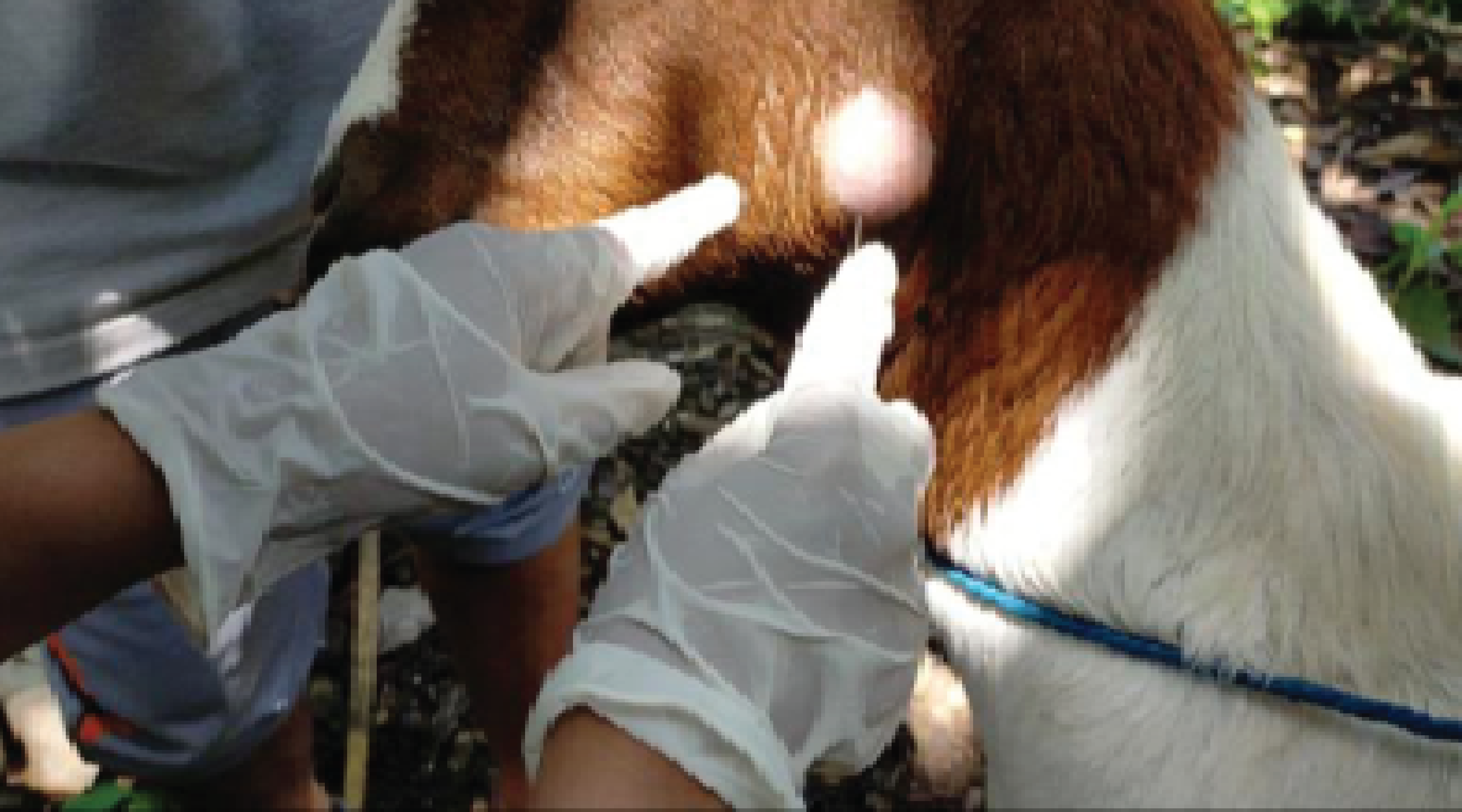
Background: An incisional hernia is a condition where the sutures on the abdominal wall rupture while the sutures on the skin remain intact. This causes abdominal viscera to protrude through the surgical incision hole, resulting in a visible lump. Purpose: Literature reports regarding incisional hernias in animals are very rarely reported; therefore, this case report aims to provide information about incisional hernias. Case: An eight month old female cat weighing 1.7 kg was presented at the Veterinary Surgery Laboratory, Veterinary Medical Faculty of Udayana University, with clinical signs of a lump on the ventral side of the abdomen, with visible stitch scars in the lump area. Upon pressing the lump, it could be reinserted, leading to a diagnosis of incisional hernia. Case Management: The cat underwent herniorrhaphy to return the visceral organs to the abdominal cavity and close the hernial opening. Post-operative care included administering cefotaxime 20 mg/kg BW intravenously twice a day for three days, followed by cefixime 5 mg/kg BW orally twice a day for four days, and non-steroidal anti-inflammatory drugs tolfenamic acid of 4 mg/kg BW subcutaneously once a day for three days. The wound area was covered with 1% framycetin sulfate tulle gauze (Daryant-Tulle®), and the stitches were removed on the 14th postoperative day. Conclusion: The wound appears dry, with well-approximated edges and no visible protrusion. It can be concluded that treatment with herniorrhaphy in this case was successful.
Abu-Seida, A.M.A., 2012. Efficacy of Diclofenac Sodium, Either Alone or Together with Cefotaxime Sodium, for Control of Postoperative Pain, in Dogs Undergoing Ovariohysterectomy. Asian Journal of Animal and Veterinary Advances, 7 (2), 180-186.
Akgül, M.B., Yildirim, O., Kiliç, S., Kaçak, K., Akgül, G., and Gülaydin, A., 2023. Right Lateral Paracosto-Abdominal Hernia In A Cat. Turkish Journal of Veterinary Research, 7 (1), 47-52.
Atiyeh, B.S., Al-Amm, C.A., El-Musa, K.A., Sawwaf, A., and Dham, R., 2003. The Effect of Moist and Moist Exposed Dressings on Healing and Barrier Function Restoration of Partial Thickness Wounds. European Journal of Plastic Surgery, 26 (1), 5-11.
Babu, M., Krishnaswamy, A., Nethra, R., and Murthy, N., 2019. Comparison of Two Different Laparotomy Techniques for Ovariohysterectomy and Post-Surgical Complications in Cats. International Journal of Current Microbiology and Applied Sciences, 8 (05), 331-334.
Bellenger, C.R., 2003. Textbook of Small Animal Surgery. Elsevier Health Sciences. Elsevier Health Sciences, 405–413.
Bolton, L.L., Monte, K., and Pirone, L.A., 2000. Moisture and Healing: Beyond the Jargon. Ostomy Wound Manage. WEF-CEP FDA project to add Approved Clinical Trial Endpoints V, 1A, 51-62.
Breuing, K., Eriksson, E., Liu, P., and Miller, D.R., 1992. Healing of Partial Thickness Porcine Skin Wounds in A Liquid Environment. Journal of Surgical Research, 52 (1), 50-58.
Carmine, A.A., Brogden, R.N., Heel, R.C., Speight, T.M., and Avery, G.S., 1983. Cefotaxime A review of Its Antibacterial Activity, Pharmacological Properties and Therapeutic Use. Drugs, 25 (3), 223-289.
Chen, W.Y.J., Rogers, A.A., and Lydon, M.J., 1992. Characterization of Biologic Properties of Wound Fluid Collected During Early Stages of Wound Healing. Journal of Investigative Dermatology, 99 (5), 559-564.
Conzemius, M.G., Brockman, D.J., King, L.G., and Perkowski, S.Z., 1994. Analgesia in Dogs After Intercostal Thoracotomy: A Clinical Trial Comparing Intravenous Buprenorphine and Interpleural Bupivacaine. Veterinary Surgery, 23 (4), 291-298.
Erwin, Karmil, T.F., Etriwati, Salim, M.N., and Rusli, A., 2021. Kesembuhan Skin Flap Rotasi Pada Kucing dengan Perawatan Dry Dressing dan Moist Dressing Secara Subjektif dan Objektif: Original Research. Acta Veterinaria Indonesiana, 9 (2), 120-126.
Fortelny, R.H., 2018. Abdominal Wall Closure in Elective Midline Laparotomy: The Current Recommendations. Frontiers in Surgery, 5 (34), 1–8.
Fossum, T.W., 2019. Small Animal Surgery. Fifth edition. Philadelphia, PA: Elsevier.
Gonzalez, A.C.D.O., Costa, T.F., Andrade, Z.D.A., and Medrado, A.R.A.P., 2016. Wound Healing - A Literature Review. Anais Brasileiros de Dermatologia, 91 (5), 614-620.
Hansen, J. and Perry, B., 1994. Helminth Parasites of Ruminants. International Laboratory for Research on Animal Diseases, 31, 1656–1664.
Kumar, B.P., Phaneendra, M., and Lakshmi, N.D., 2017. Surgical Management of Perineal Hernia Associated With Inguinal Hernia in A Spitz. Journal of Entomology and Zoology Studies, 5 (3), 902-904.
Lima, A.J.S., Lima, W.C., Lima, D.A.S.D., Sala, P.L., Borges, T.B., and Quessada, A.M., 2021. Incisional Hernia in a Dog. Acta Scientiae Veterinariae, 49 (1), 1-5.
Mandara, I. and Pemayun, I.G.A.G.P., 2021. Laporan Kasus: Penanganan Hernia Paracostalis Pada Kucing Lokal Jantan dengan Laparotomi. Indonesia Medicus Veterinus, 10 (4), 644-656.
Mazzocchi, M., Dessy, L.A., Ranno, R., Carlesimo, B., and Rubino, C., 2011. "Component Separation” Technique and Panniculectomy for Repair of Incisional Hernia. The American Journal of Surgery, 201 (6), 776-783.
Morrison, B., Christensen, S., Yuan, W., Brown, J., Amlani, S., and Seidenberg, B., 1999. Analgesic Efficacy of The Cyclooxygenase-2-specific Inhibitor Rofecoxib in Post-Dental Surgery Pain: A Randomized, Controlled Trial. Clinical Therapeutics, 21 (6), 943–953.
Muraro, L. and White, R.S., 2014. Complications of Ovariohysterectomy Procedures Performed in 1880 Dogs. Tierärztliche Praxis Ausgabe K: Kleintiere / Heimtiere, 42 (05), 297-302.
Rabbani, A.H., Khan, Y.R., Naseer, O., Safwan, H.M., Waheed, A., Ali, A., and Waqas, M., 2021. Biocompatibility and Postoperative Complications of Different Subcutaneous Suturing Methods in Cats Undergoing Ovariohysterectomy by Midline Incision. The Thai Journal of Veterinary Medicine., 3 (51), 577-585.
Rizk, A. and Samy, A., 2016. Diagnosis and Surgical Repair of Entero-Cystocele in A Cat. Open Veterinary Journal, 6 (3), 162-164.
Sattasathuchana, P., Phuwapattanachart, P., and Thengchaisri, N., 2018. Comparison of Post-Operative Analgesic Efficacy of Tolfenamic Acid and Robenacoxib in Ovariohysterectomized Cats. Journal of Veterinary Medical Science, 80 (6), 989-996.
Sewoyo, P.S., Widyastuti, S.K., Erawan, I.G.M.K., 2022. Laporan Kasus: Keberhasilan Penanganan Rinitis Unilateral Kronis yang Menyebabkan Polisitemia Sekunder pada Kucing Lokal. Indonesia Medicus Veterinus, 11 (6), 898-911
Slingsby, L.S. and Waterman"Pearson, A.E., 2000. Postoperative Analgesia in The Cat After Ovariohysterectomy by Use of Carprofen, Ketoprofen, Meloxicam or Tolfenamic Acid. Journal of Small Animal Practice, 41 (10), 447–450.
Taddio, A., Katz, J., Ilersich, A.L., and Koren, G., 1997. Effect of Neonatal Circumcision on Pain Response During Subsequent Routine Vaccination. The Lancet, 349 (9052), 599-603.
Travis, B.M., Hayes, G.M., Vissio, K., Harvey, H.J., Flanders, J.A., and Sumner, J.P., 2018. A Quilting Subcutaneous Suture Pattern to Reduce Seroma Formation and Pain 24 Hours After Midline Celiotomy in Dogs: A Randomized Controlled Trial. Veterinary Surgery, 47 (2), 204-211.
Tsugawa, A.J. and Verstraete, F.J.M., 2020. Suture Materials and Biomaterials. In: Oral and Maxillofacial Surgery in Dogs and Cats. Elsevier, 79-91.
Wallace, H.A., Basehore, B.M., and Zito, P.M., 2022. Wound Healing Phases. StatPearls Publishing.
William, W. and Faiz Tuma, 2023. Incisional Hernia. National Center for Biotechnology Information.
Copyright (c) 2023 Palagan Senopati Sewoyo, Made Santi Purwitasari, Pramita Rabiulfa, I Wayan Wirata

This work is licensed under a Creative Commons Attribution-ShareAlike 4.0 International License.
- The journal allows the author to hold the copyright of the article without restrictions.
- The journal allows the author(s) to retain publishing rights without restrictions.
- The legal formal aspect of journal publication accessibility refers to Creative Commons Attribution Share-Alike (CC BY-SA).

Journal of Applied Veterinary Science and Technology is licensed under a Creative Commons Attribution-ShareAlike 4.0 International License