Fascioliasis: A Zoonotic Disease and Diagnostic Capture Using Radiological Imaging
Downloads
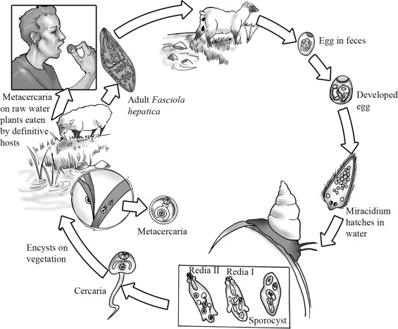
Fascioliasis, also known as hepatic distomatosis or fasciolosis, is a zoonotic infection caused by the trematodes of Fasciola. The usual reservoir for this parasitic disease is herbivorous mammals, including humans, sheep, goats, and cattle. However, humans can contract this zoonosis infection by ingesting metacercaria, a juvenile trematode stage, which adheres to aquatic vegetation. Fascioliasis is typically present asymptomatically. However, human fascioliasis may have symptoms such as eosinophilia, abdominal discomfort, and various corroborative findings covering multiple diagnostic modalities. These diagnostic options include parasitological fecal examination, which observes the parasite in the feces; radiological imaging techniques, which envision the anatomical abnormalities created by the invasion; and serological studies, which could detect the immune response system to the infestation of the parasite. This review article aims to characterize fascioliasis in terms of zoonotic occurrence, outline the available diagnostic modalities, and highlight the specific significance of radiological imaging. This may contribute to the timely and adequate identification of the condition. This review article may contribute to forming the professional dialogue concerning fascioliasis, including its epidemiology, clinical presentation, and differential diagnostics
Aftab, A. et al. (2024) ’Advances in diagnostic approaches to Fasciola infection in animals and humans: An overview,’ Journal of Helminthology, 98, e12. Available at: https://doi.org/10.1017/S0022149X23000950
Behar, J.M., Winston, J.S. and Borgstein, R. (2009) ’Hepatic fascioliasis at a London hospital - The importance of recognising typical radiological features to avoid a delay in diagnosis,’ British Journal of Radiology, 82(981), pp. 189-193. Available at: https://doi.org/10.1259/bjr/78759407
Bogitsh, B.J., Carter, C.E. and Oeltmann, T.N. (2012). Human Parasitology. 4th edition. Academic Press, Elsevier Inc. Available at: https://doi.org/10.1016/C2010-0-65681-1
Dusak, A. et al. (2012) ‘Radiological Imaging Features of Fasciola hepatica Infection - A Pictorial Review,’ Journal of Clinical Imaging Science, 2(2), Available at: https://doi.org/10.4103/2156-7514.92372
Fang, W. et al. (2022) ‘Exploration of animal models to study the life cycle of Fasciola hepatica,’ Parasitology, 149(10), pp. 1349-1355. Available at: https://doi.org/10.1017/S0031182022000609
Farrar, J. et al. (2014). Manson’s Tropical Infectious Diseases. 23rd edition. Saunders Ltd. Copyright Elsevier Ltd. Available at: https://doi.org/10.1016/C2010-0-66223-7
Keiser, J. and Utzinger, J. (2005) ‘Emerging foodborne Trematodiasis,’ Emerging Infectious Diseases, 11(10), pp. 1507-1514. Available at: https://doi.org/10.3201/eid1110.050614
Lalor, R. et al. (2021) ‘Pathogenicity and virulence of the liver flukes Fasciola hepatica and Fasciola Gigantica that cause the zoonosis Fasciolosis,’ Virulence, 12(1), pp. 2839-2867. https://doi.org/10.1080/21505594.2021.1996520.
Mas-Coma, S., Bargues, M.D. and Valero, M.A. (2014) ‘Diagnosis of human fascioliasis by stool and blood techniques: update for the present global scenario,’ Parasitology, 141(14), pp.1918-1946. Available at: https://doi.org/10.1017/S0031182014000869.
Nyindo M. and Lukambagire, A.H. (2015) ‘Fascioliasis: An Ongoing Zoonotic Trematode Infection,’ BioMed Research International, (article ID 786195), pp. 1-8. Available at: https://doi.org/10.1155/2015/786195.
Prasetyo, D.A. et al. (2023). ‘High prevalence of liver fluke infestation, Fasciola gigantica, among slaughtered cattle in Boyolali District, Central Java,’ Open Veterinary Journal, 13(5), pp. 654-662. Available at: https://doi.org/10.5455/OVJ.2023.V13.I5.19.
Preza, O. et al. (2019) ‘Fascioliasis: A challenging differential diagnosis for radiologists,’ The Journal of Radiology Case Reports, 13(1), pp. 11-16. Available at: https://doi.org/10.3941/jrcr.v13i1.3451.
Rinca, K.F. et al. (2019) ‘Trematodiasis occurrence in cattle along the Progo River, Yogyakarta, Indonesia,’ Veterinary World, 12(4), pp. 593-597. Available at: https://doi.org/10.14202/vetworld.2019.593-597.
Salahshour, F. and Tajmalzai, A. (2021) ‘Imaging findings of human hepatic fascioliasis: a case report and review of the literature.’ Journal of Medical Case Reports, 15(324), pp. 1-6. Available at: https://doi.org/10.1186/s13256-021-02945-9.
Taylor, M.A., Coop, R.L. and Wall, R.L. (2016). Veterinary Parasitology. 4th edition. Willey Blackwell. Available at: https://doi.org/10.1016/s0167-5877(97)00068-8.
Tenorio, J. C. B. and Molina, E. C. (2021) ‘Monsters in our food: Foodborne trematodiasis in the Philippines and beyond,’ Veterinary Integrative Sciences, 19(3), 467-485. Available at: https://doi.org/10.12982/vis.2021.038.
World Health Organization (2008). Fact sheet on fascioliasis. In Action Against Worms. Newsletter, 10, pp. 1–8. World Health Organization, Geneva, Switzerland.
Copyright (c) 2024 Journal of Parasite Science (JoPS)

This work is licensed under a Creative Commons Attribution-NonCommercial-ShareAlike 4.0 International License.
- Every manuscript submitted to must observe the policy and terms set by the Journal of Parasite Science
- Publication rights to manuscript content published by the Journal of Parasite Science is owned by the Journal of Parasite Science with the consent and approval of the author(s) concerned
- Authors and other parties are bound to the Creative Commons Attribution-NonCommercial-ShareAlike 4.0 International License for the published articles, legal formal aspect of journal publication accessibility refers to Creative Commons Attribution-NonCommercial-ShareAlike 4.0 International License (CC BY-NC-SA)
- By submitting the manuscript, the author agrees to the requirement that the copyright of the submitted article will be transferred to Journal of Parasite Science as the publisher of the journal. The intended copyright includes the right to publish articles in various forms (including reprints). journal of parasite science retains the publishing rights to published articles.


