A CASE REPORT: ENHANCING TARSAL AND MUSCULAR SUPPORT FOR ECTROPION CORRECTION IN TESSIER 3 AND 5 FACIAL CLEFT

Downloads
Highlights:
- The oblique facial cleft is an uncommon and intricate craniofacial anomaly characterized by a low occurrence rate and significant variability.
- The treatment is intricate and relies on the surgeon's expertise and discernment.
- Utilizing a combination of Tarsal Strip and Midface Lifting techniques could serve as an alternative approach to address ectropion by enhancing both tarsal and muscular support.
Abstract:
Introduction: Facial cleft is a rare and challenging craniofacial malformation with a low incidence ranging from 0.75 to 5.4 per 1000 common cleft. Due to the high variance in its occurrence, good techniques have not been established yet.
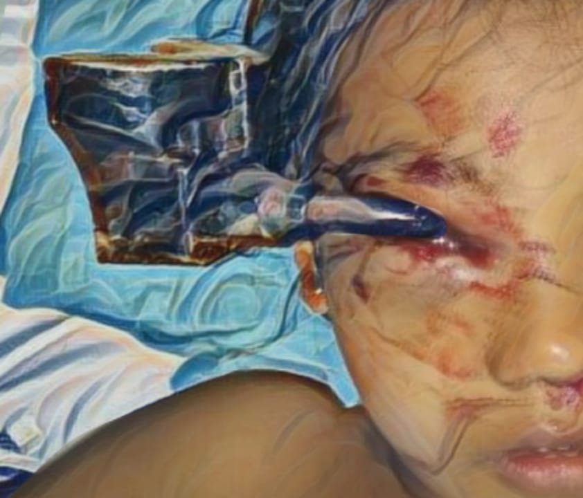
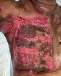
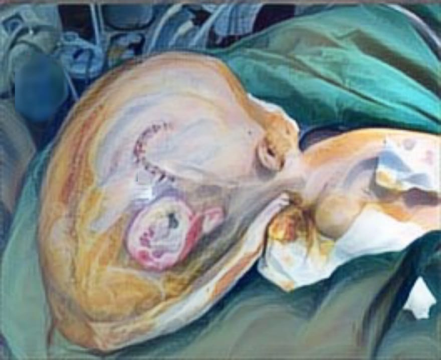
Case Illustration: In this case study, we present the medical history of a 45-year-old woman who previously underwent multiple surgeries for the reconstruction of her lower eyelid. The initial surgeries were performed to address a unilateral facial cleft classified as Tessier 3 and 5, utilizing ear cartilage grafts for the tarsal plate. However, she subsequently experienced complications, including ectropion, epiphora, and soft tissue deformities around her left eye. The goal of the current surgical intervention is to rectify the ectropion and enhance overall facial aesthetics for improved outcomes.
Discussion: The primary objectives of this surgical techniques are to enhance the tarsal support through the tarsal strip technique and to provide muscle support using the mid-face lifting technique. Additionally, we removed the scar and excess tissue from the lower lid to adjust the tarsal's proper length. These methods aim to address the ectropion and prevent its recurrence.
Conclusion: This combination of techniques can be a potential alternative for rectifying ectropion by reinforcing both tarsal and muscular support structures.
MOHD Ashraf Darzi MS, MC, Nisar Ahmad Chowdri MS. Oblique Facial Cleft_ A report of Tessier Number 3,4,5 and 9 Cleft [Internet]. [cited 2023 Jul 23]. Available from: https://pubmed. ncbi.nlm.nih.gov/8399272/
J. C. H. Van der Meulen MD. Oblique Facial Clefts : Pathology Etiology and Reconstruction .pdf. Plastic and Reconstrucive Surgery. 1985;75(2):212 –24.
Tessier P. Anatomical Classification of Facial, Cranio-Facial and Latero-Facial Clefts*. J max-fac Surg [Internet]. 1976 [cited 2023 Aug 1];4(2):69–92. Available from: https://pubmed.ncbi. nlm.nih.gov/820824/
Budihardja AS, Lutfianto B, Liman NP, Hiesmantjaja H, Wolff KD. Rare Facial Cleft: Surgical Treatment and Middle-Term Follow-up During Charity Operation. Craniomaxillofac Trauma Reconstr. 2020.13(2):138–42.
Freitas R da S, Alonso N, Shin JH, Busato L, Dall' R, Tolazzi O, et al. The Tessier Number 5 Facial Cleft: Surgical Strategies and Outcomes in Six Patients. Cleft Palate-Craniofacial Journal. 2009;46(2):179–86.
Balaji S. Two-stage corrections of rare facial tessier's cleft - 3,4,5,6,7. Ann Maxillofac Surg [Internet]. 2017 [cited 2023 Oct 11];7(2):287. Available from: https://www.ncbi.nlm.nih.gov/pmc/articles/PMC5717909/pdf/AMS-7-287.
Jue MS, Yoo J, Kim MS, Park HJ. The lateral tarsal strip for paralytic ectropion in patients with leprosy. Ann Dermatol. 2017.29(6):742–6.
Mendelson BC. Fat Preservation Technique of Lower-lid Blepharoplasty. Aesthet Surg J. 2001;21(5):45–459.
Lee TY, Cha JH, Ko HW. Rejuvenation of the Lower Eyelid and Midface with Deep Nasolabial Fat Lift in East Asians. Plast Reconstr Surg. 2023.151(6):931E-940E.
Anderson RL, Gordy DD. The Tarsal Strip Procedure. Arch Ophthalmol [Internet]. 1979;97:2192–6. Available from: http://archopht.jamanetwork. com/
Sari E. Tessier number 30 facial cleft: a rare maxillofacial anomaly. Turk J Plast Surg. 2018;26(1):12–19. doi: 10.4103/tjps.tjps_7_18.
Kalantar-Hormozi, Abdoljalil MD*; Abbaszadeh-Kasbi, Ali MD" ; Goravanchi, Farhood MD"¡; Davai, Nazanin Rita MD§. Prevalence of Rare Craniofacial Clefts. Journal of Craniofacial Surgery.2017.28(5):p e467-e470. doi:10.1097/SCS.0000000000003771
Palukuri L, JSR, DVN Tessier 30 Facial Cleft: A Rare Craniofacial Anomaly. Int J Clin Pediatr Dent 2023;16(1):177-179.
David DJ, Moore MH, Cooter RD, Chow SK. The Tessier number 9 cleft. Plast Reconstr Surg.1989.83:520–7.
Bagheri, Abbas et al. Tessier Number 9 Craniofacial Cleft Associated with Goldenhar Syndrome and Its Surgical Management: A Report of a Rare Case. Journal of Current Ophthalmology. 2023. 35(1):p 93-95. doi: 10.4103 /joco.joco_153_22
Kawamoto HK Jr. The kaleidoscopic world of rare craniofacial clefts:Order out of chaos (Tessier classification). Clin Plast Surg. 1976;3:529–72.
Dumortier R, Delhemmes P, Pellerin P. Bilateral Tessier No. 9 cleft. J Craniofac Surg. 1999;10:523–5.
Bubanale SC, Kurbet SB, De Piedade Sequeira LM. A rare case of cleft number nine associated with atypical cleft number two. Indian J Ophthalmol. 2017;65:610–2.
Tessier P. Anatomical classification facial, cranio-facial and latero-facial clefts. J Maxillofac Surg. 1976;4:69–92.
Afifi et al.Long-term follow-up of a Tessier number 5 facial cleft Craniomaxillofac Trauma Reconstr ,2011. 4(1):35-42
Wolfe et al.Commentary on: Afifi A.M., Djohan R., Sweeney W., Brooks S., Connolly J., et al. Long-term follow-up of a Tessier number 5 facial cleft Craniomaxillofac Trauma Reconstr, 2011.4(1):40-42.
Chattopadhyay et al.Bilateral Tessier number 5 facial cleft with limb constriction ring: the first case report with an update of literature review. J Oral Maxillofac Surg Med Pathol, 2013.25(1):43-45.
Ramanathan et al., A rare case of multiple oblique facial clefts with supernumerary teeth: case report.Craniomaxillofac Trauma Reconstr,2012.5(4):239-242.
Abdollahifakhim et al. A bilateral Tessier number 4 and 5 facial cleft and surgical strategy: a case report.Iran J Otorhinolaryngol,2013.25(73):259-262
Racz, C., (2018). Phenotypic spectrum of Tessier facial cleft number 5. Journal of Cranio-Maxillofacial Surgery, 2018. 46 (1):22-27.
Abbe R. A new plastic operation for the relief of deformity due to double harelip. Plast Reconstr Surg.1968.42(5):481–483.
Van Slyke AC, et al.The Anatomical Subunit Approach to Managing Tessier Numbers 3 and 4 Craniofacial Clefts. Plast Reconstr Surg Glob Open. 2022.10(9):e4553. doi: 10.1097/GOX. 0000000000004553.
Mishra, R. K., & Purwar, R. Formatting the surgical management of Tessier cleft types 3 and 4. Indian Journal of Plastic Surgery, 2009.42(S 01), S174-S183.
David DJ, Fracs F. New perspectives in the management of severe cranio-facial deformity*. In: Annals of the Royal College of Surgeons of England [Internet]. Adelaide; 1984 [cited 2023 Sep 15]. p. 270–9. Available from:https://pubmed.ncbi.nlm.nih.gov/6742741/
Soedjana H, Prasetyo AT, Dewi C. Paranasal transposition flap in facial soft tissue reconstruction of facial cleft Tessier type 3 & ADAM complex: A case report. Int J Surg Case Rep. 2021.1;87.
Wenbin Z, Hanjiang W, Xiaoli C, Zhonglin L. Tessier 3 Cleft With Clinical Anophthalmia: Two Case Reports and a Review of the Literature. Cleft Palate–Craniofacial Journal [Internet]. 2007 Jan [cited 2023 Aug 9];44(1):102–5. Available from: https://pubmed.ncbi .nlm.nih.gov/17214533/
Roddi R, Oo A, Pepe E, Naing E, Sung S. Surgical Strategy for the Treatment of Facial Clefts. Surgical Techniques Development [Internet]. 2023 Jan 25 [cited 2023 Oct 16];12(1):34–42. Available from: https://www.mdpi. com/2038-9582/ 12/1/2
Menard RM, Moore MH, David DJ. Tissue Expansion In Reconstruction of Tessier Craniofacial Cleft_ Aseries of 17 Patients. Plast Reconstr Surg. 1999;103(3):779–86.
Copyright (c) 2023 Teuku Adifitrian, Hendra Tri Hartono, Agustina Indah Garnasih

This work is licensed under a Creative Commons Attribution-ShareAlike 4.0 International License.
JURNAL REKONSTRUKSI DAN ESTETIK by Unair is licensed under a Creative Commons Attribution-ShareAlike 4.0 International License.
- The journal allows the author to hold copyright of the article without restriction
- The journal allows the author(s) to retain publishing rights without restrictions.
- The legal formal aspect of journal publication accessbility refers to Creative Commons Attribution Share-Alike (CC BY-SA)


