IMPLEMENTATION OF AN OCCLUSAL WAFER IN SEVERE MANDIBULAR FRACTURE CASES WITH POST-ORIF MALOCCLUSION: A CASE SERIES
Downloads
Highlights:
- This study shows that occlusal wafers can effectively correct malocclusion in patients with segmental mandibular fractures after ORIF plating.
- Occlusal wafers help reshape the dental arch within 2 to 4 weeks, reduce surgery time, and simplify follow-up care, making them a valuable option for surgeons.
Abstract:
Introduction: Improper treatment of severe mandibular fractures can lead to malocclusion, which poses a significant challenge for reconstructive surgeons. The occlusal wafer provides an effective solution for managing malocclusion following ORIF plating of maxillofacial fractures during the one-month postoperative evaluation period. Made from acrylic resin, the occlusal wafer serves as an intermediate guide in orthognathic surgery. It helps reposition the maxilla, adjust the mandible, and modify the jawbones to achieve ideal occlusion. The device can reshape the dental arch to any pre-planned position within 2 to 4 weeks.
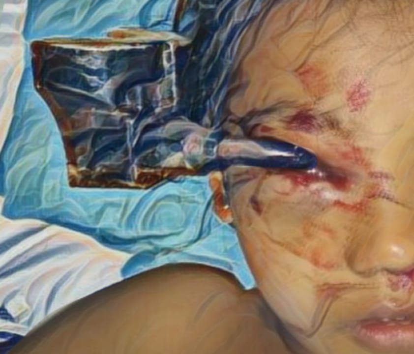
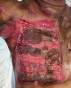
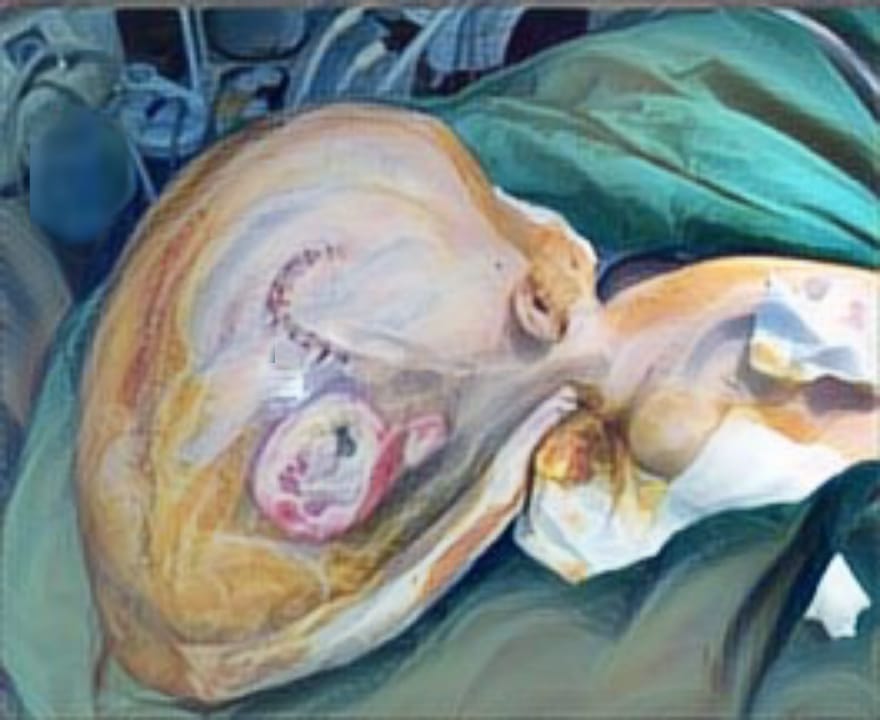
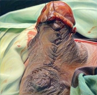
Case Illustration: We present two cases of patients with segmental fractures.Case 1: A 26-year-old male also had segmental fractures of the left angle and right body of the mandible. He achieved occlusion after ORIF plating; however, malocclusion developed during the three-week follow-up. Case 2: A 28-year-old female presented with segmental fractures of the left angle and right body of the mandible. She initially achieved occlusion after ORIF plating, but malocclusion was noted during the one-month follow-up.
Discussion: Both of these patients had segmental fractures and experienced malocclusion following ORIF plating, but occlusion was achieved after occlusal wafer installation.
Conclusion: The use of an occlusal wafer facilitates optimal occlusion, streamlines the surgical procedure by reducing operating time, and enhances the ease of postoperative monitoring. This approach proves particularly valuable in cases where ORIF plating has been performed yet ideal occlusal alignment remains unachieved.
Singaram M & Udhayakumar RK. Prevalence, pattern, etiology, and management of maxillofacial trauma in a developing country: a retrospective study. Journal of the Korean Association of Oral and Maxillofacial Surgeons, 2016; 42(4), 174-181. DOI:10.5125/jkaoms.2016.42.4. 174
Vega LG. Reoperative mandibular trauma: management of posttraumatic mandibular deformities. Oral and Maxillofacial Surgery Clinics, 2011; 23(1):47-61. DOI:10.1016/ j.coms.2010.12.003
Motamedi MHK. An assessment of maxillofacial fractures: a 5-year study of 237 patients. Journal of Oral and Maxillofacial Surgery, 2003; 61(1):61-64. DOI:10.1053/joms.2003.50049
Fordyce AM, Lalani Z, Songra AK, Hildreth AJ, Carton ATM, & Hawkesford JE. Intermaxillary fixation is not usually necessary to reduce mandibular fractures. British Journal of Oral and Maxillofacial Surgery, 1999; 37(1):52-57. DOI:10.105 4/bjom.1998.0372
Dawson PE. New definition for relating occlusion to varying conditions of the temporomandibular joint. The Journal of prosthetic dentistry,1995; 74(6): 619-627. DOI:10.1016/S0022-3913(05)80315-4
Imola MJ, Ducic Y, & Adelson RT. The Secondary Correction of Post‐Traumatic Craniofacial Deformities. Otolaryngology–Head and Neck Surgery, 2008; 139(5), 654-660.DOI:10.1016/j.otohns.2008.07.031
Lindorf, H. H., & Steinhäuser, E. W. Correction of jaw deformities involving simultaneous osteotomy of the mandible and maxilla. Journal of maxillofacial surgery, 1978; 6:239-244. DOI:10.1016/ S0301-0503(78)80099-X
Der‐Martirosian C, Gironda MW, Black EE, Belin TR, & Atchison KA. Predictors of treatment preference for mandibular fracture. Journal of public health dentistry, 2010;70(1):13-18. DOI:10.1111/j.1752-7325.2009.00138.x
Naeem A, Gemal H, Reed D. Imaging in traumatic mandibular fractures. Quantitative imaging in medicine and surgery,2017;7(4):469–479. DOI: 10.21037/qims.2017.08.06
Nardi C, Vignoli C, Pietragalla, M. et al. Imaging of mandibular fractures: a pictorial review. Insights Imaging,2020; 11(1):30. DOI: 10.1186/s13244-020-0837-0
Marker P, Nielsen A, Bastian HL.Fractures of the mandibular condyle. Part 1: patterns of distribution of types and causes of fractures in 348 patients. British journal of oral and maxillofacial surgery,2000; 38(5), 417-421.DOI:10.1054/bjom.2000.0317
Munante-Cardenas JL, Nunes PHF, & Passeri LA. Etiology, treatment, and complications of mandibular fractures. Journal of Craniofacial Surgery, 2015; 26(3): 611-615. DOI: 10.1097/SCS. 0000000000001273
Anyanechi CE & Saheeb BD. Mandibular sites prone to fracture: analysis of 174 cases in a Nigerian tertiary hospital. Ghana medical journal, 2011;45(3):111-114. Available at: https://www.ajol.info/index. php/gmj/article/view/74317
Boffano P, Kommers SC, Karagozoglu KH, Gallesio C, & Forouzanfar T. Mandibular trauma: a two-centre study. International journal of oral and maxillofacial surgery, 2015; 44(8): 998-1004. DOI: 10.1016/ j.ijom.2015.02.022
Jabaz D. Mandible fracture. StatPearls. 2022;1–7.
Kim SY, Choi YH, & Kim K. Postoperative malocclusion after maxillofacial fracture management: a retrospective case study. Maxillofacial plastic and reconstructive surgery,2018; 40(27):1-8. DOI:10.1186/ s40902-018-0167-z
Nakamura S, Takenoshita Y & Oka M. Complications of miniplate osteosynthesis for mandibular fractures. Journal of oral and maxillofacial surgery, 1994; 52(3): 233-238. DOI:10.1016/0278-2391(94)9 0289-5
Maron G, Kuhmichel A, Schreiber G. Secondary Treatment of Malocclusion/Malunion Secondary to Condylar Fractures. Atlas Oral Maxillofac Surg Clin North Am [Internet]. 2017;25(1):47–54. DOI: 10.1016/j.cxom. 2016.10.003
Bamber MA & Harris M. The role of the occlusal wafer in orthognathic surgery; a comparison of thick and thin intermediate osteotomy wafers. Journal of cranio-maxillofacial surgery,1995; 23(6):396-400. DOI: 10.1016/S1010-5182(05)80137-4
Vachircmon A, Bamber MA, Suwantawakup A, & Nitiwaragkul W. Review of Orthognathic Surgery Occlusal Wafers Application in 185 Patients. The Thai Journal of Surgery, 2002; 23(1):21-26. Available at: https://he02.tcithaijo.org/index.php/ThaiJSurg/article/view/243117/165309
Bocca AP, Kittur MA, Gibbons AJ, & Sugar AW. A new type of occlusal wafer for orthognathic surgery. British Journal of Oral and Maxillofacial Surgery, 2002; 40(2):151-152. DOI:10.1054/bjom.2001.0760
Bamber MA & Vachiramon A. Surgical wafers: a comparative study. Journal of Contemporary Dental Practice, 2005; 6(2): 99-106. Available at: https://www.thejcdp.com/doi/JCDP/pdf/10.5005/jcdp-6-2-99
Proffit W, White P. Surgical orthodontic treatment. First. Toronto, Chicago: Mosby; 1991. 203 p.
Rehman KU, Shetty S, Dover S. Simple way to secure an occlusal wafer in patients with compromised dentition. British Journal of Oral and Maxillofacial Surgery, 2008;46(6):509. DOI: 10.1016/j.bjoms. 2008.02.011
Pearl EE, Joevitson M, Sreelal T, Chandramohan G, Mohan A, Hines AJ. Marking the invisible – A review of the various occlusal indicators and techniques. International Journal of Applied Dental Sciences, 2020;6(2):377–81. Available at: https://www.oraljournal.com/archives/2020/6/2/F/6-2-48
Copyright (c) 2024 Chintia Amelia Pratiwi, Mirnasari Amirsyah, Teuku Nanda Putra

This work is licensed under a Creative Commons Attribution-ShareAlike 4.0 International License.
JURNAL REKONSTRUKSI DAN ESTETIK by Unair is licensed under a Creative Commons Attribution-ShareAlike 4.0 International License.
- The journal allows the author to hold copyright of the article without restriction
- The journal allows the author(s) to retain publishing rights without restrictions.
- The legal formal aspect of journal publication accessbility refers to Creative Commons Attribution Share-Alike (CC BY-SA)


