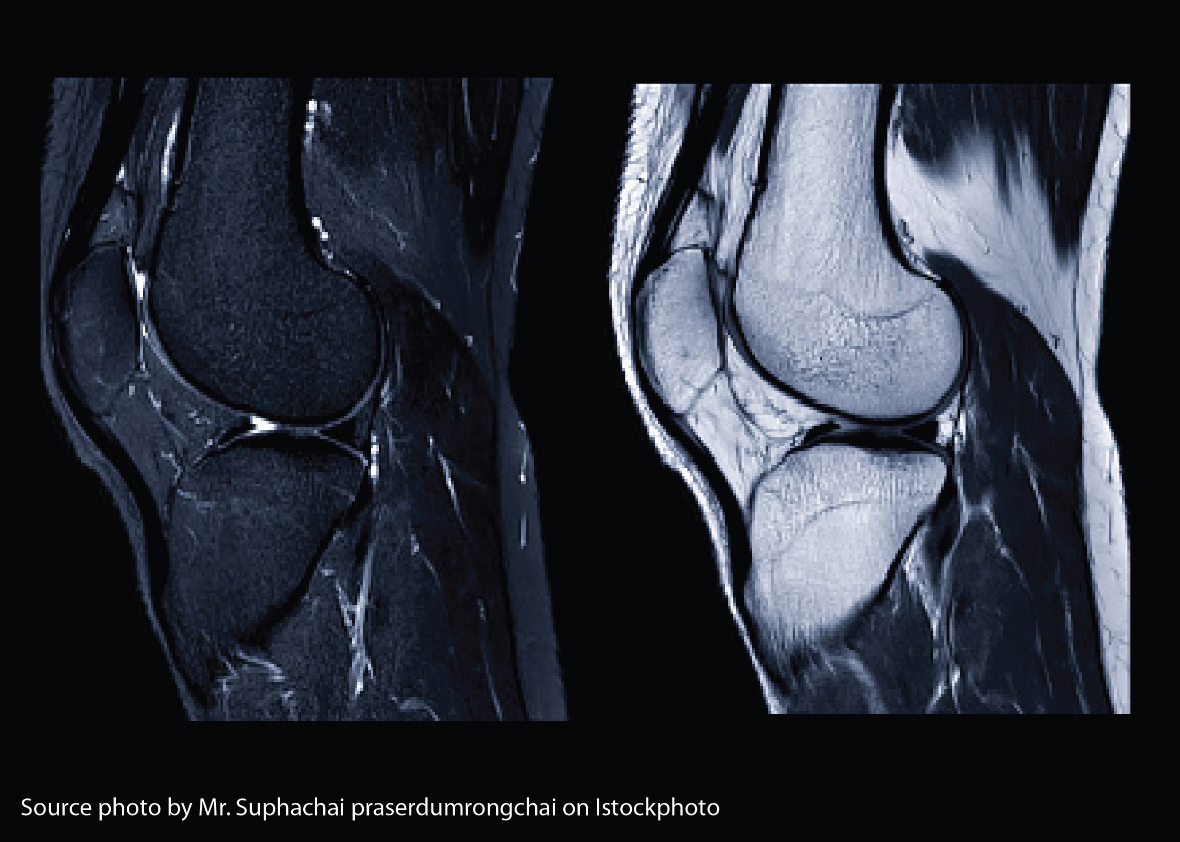
DIFFERENCE OF ANATOMY INFORMATION MRI KNEE JOINT ON VARIATION OF TIME REPETITION SEQUENCES STIR IN SAGITTAL SLICES

Downloads
Background: Time Repetition (TR) is one of the main parameters of Inversion Recovery. Purpose: The purpose of this study to determine differences in anatomical MRI information on the variation of the knee joint TR sequences STIR Sagittal slices.Method:Type of research is experimental. The study was conducted with MRI 1.5 Tesla. Data in the form of 42 image sequences STIR MRI knee joint with TR 3500, 4000, 4500, 5000, 5500, 6000, and 6500 ms. Anatomical assessments on the anterior cruciate ligament, posterior cruciate ligament, articular cartilage, and meniscus were performed by a radiologist. Data analyzed by Friedman and Wilcoxon test.Result:The results showed that there were differences in the MRI anatomical information of the knee joint of the STIR sagitas slice in the TR variation with p-value <0.001. There is a difference in anatomical information between TR 5000 and 6000 ms (p-value = 0.034), TR 5000 and 6500 ms(p-value = 0.024), TR 5500 and 6500 ms (p-value = 0.038). There is no difference in anatomical information between TR 4500 and 5000 ms (p-value = 0.395), TR 4500 and 5500 ms (p-value = 0.131), TR 4500 and 6000 ms (p-value = 0.078), TR 4500 and 6500 ms (p-value = 0.066), TR 5000 and 5500 ms (p-value = 0.414), TR 5500 and 6000 ms (p-value = 0.102), TR 6000 and 6500 ms (p-value = 0.083). Conclusion:The optimal value to produce anatomical information of the knee joint sagittal MRI sequences STIR is TR 4500 ms.
Bitar, R., Leung, G., Perng, R., Tadros, S., Moody, A.R., Sarrazin, J., McGregor, C., Christakis, M., Symons, S., Nelson, A., Roberts, T.P., 2006. MRI Pulse Sequences What Every Radiologist Wants to Know but Is Afraid to Ask. Radiographics 26, 513–537.
Freitas, A., 2016. Musculoskeletal MRI [WWW Document]. URL http://www.freitasrad.net/pages/Basic_MSK_MRI/Knee.htm. (accessed 9.5.19).
McRobbie, D.W., Moore, E.A., Graves, M.J., Prince, M.R., 2006. MRI From Picture to Proton, Second Edition. New York. Cambridge University Press, New York.
Moeller, T.B., Reif, E., 2003. MRI Parameters and Positioning. In: Radiology. p. 366.
Notosiswoyo, M., Suswaty, S., 2004. Artikel Pemanfaatan Magnetic Resonance Imaging (MRI) Sebagai Sarana Diagnosa Pasien. Media Penelit. dan Pengemb. Kesehat. 14.
R., A., V., J., 2013. Osteoarthritis of the Knee Joint - An Overview. J. Indian Acad. Clin. Med. 14, 154–162.
Westbrook, C., Roth, C.K., Talbot, J., 2011. MRI In Practice, 4 th Editi. ed. John Wiley and Sons Ltd, Chicester, United Kingdom.
Wu, J., Lu, L.-Q., Gu, J.-P., Yin, X.-D., 2012. The Application of Fat-Suppression MR Pulse Sequence in the Diagnosis of Bone-Joint Disease. Clin. Eng. Radiat. Oncol. 1, 88–94.
Copyright (c) 2020 Journal of Vocational Health Studies

This work is licensed under a Creative Commons Attribution-NonCommercial-ShareAlike 4.0 International License.
- The authors agree to transfer the transfer copyright of the article to the Journal of Vocational Health Studies (JVHS) effective if and when the paper is accepted for publication.
- Legal formal aspect of journal publication accessibility refers to Creative Commons Attribution-NonCommercial-ShareAlike (CC BY-NC-SA), implies that publication can be used for non-commercial purposes in its original form.
- Every publications (printed/electronic) are open access for educational purposes, research, and library. Other that the aims mentioned above, editorial board is not responsible for copyright violation.
Journal of Vocational Health Studies is licensed under a Creative Commons Attribution-NonCommercial-ShareAlike 4.0 International License


