COMPARISON MEASUREMENT AND CALCULATION OF BRAIN TUMOR IN MRI MODALITIES UTILISING SPIN-ECHO PULSE SEQUENCE T1-WEIGHTED CONTRAST AND DIGITAL IMAGE PROCESSING APPLICATIONS

Downloads
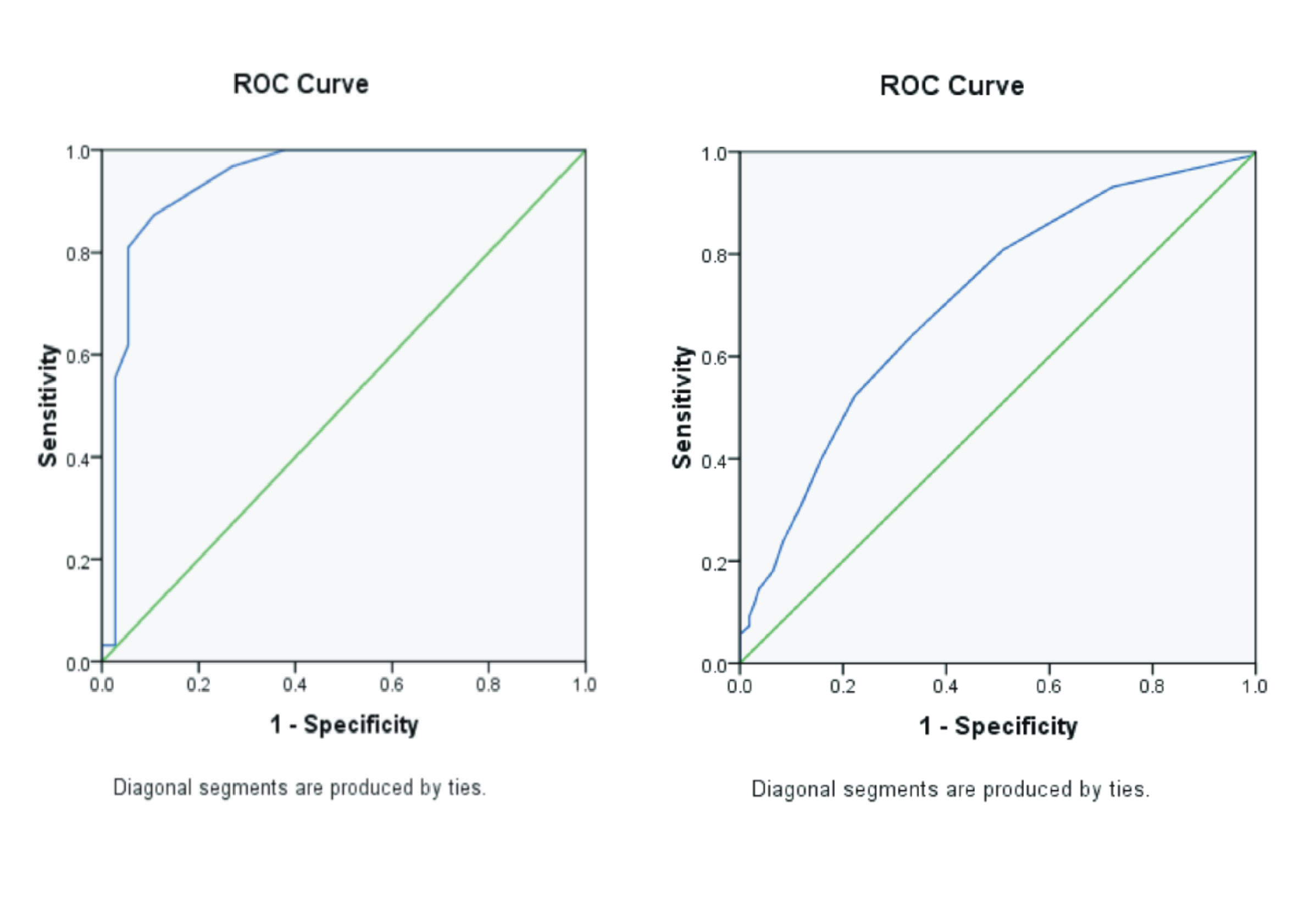
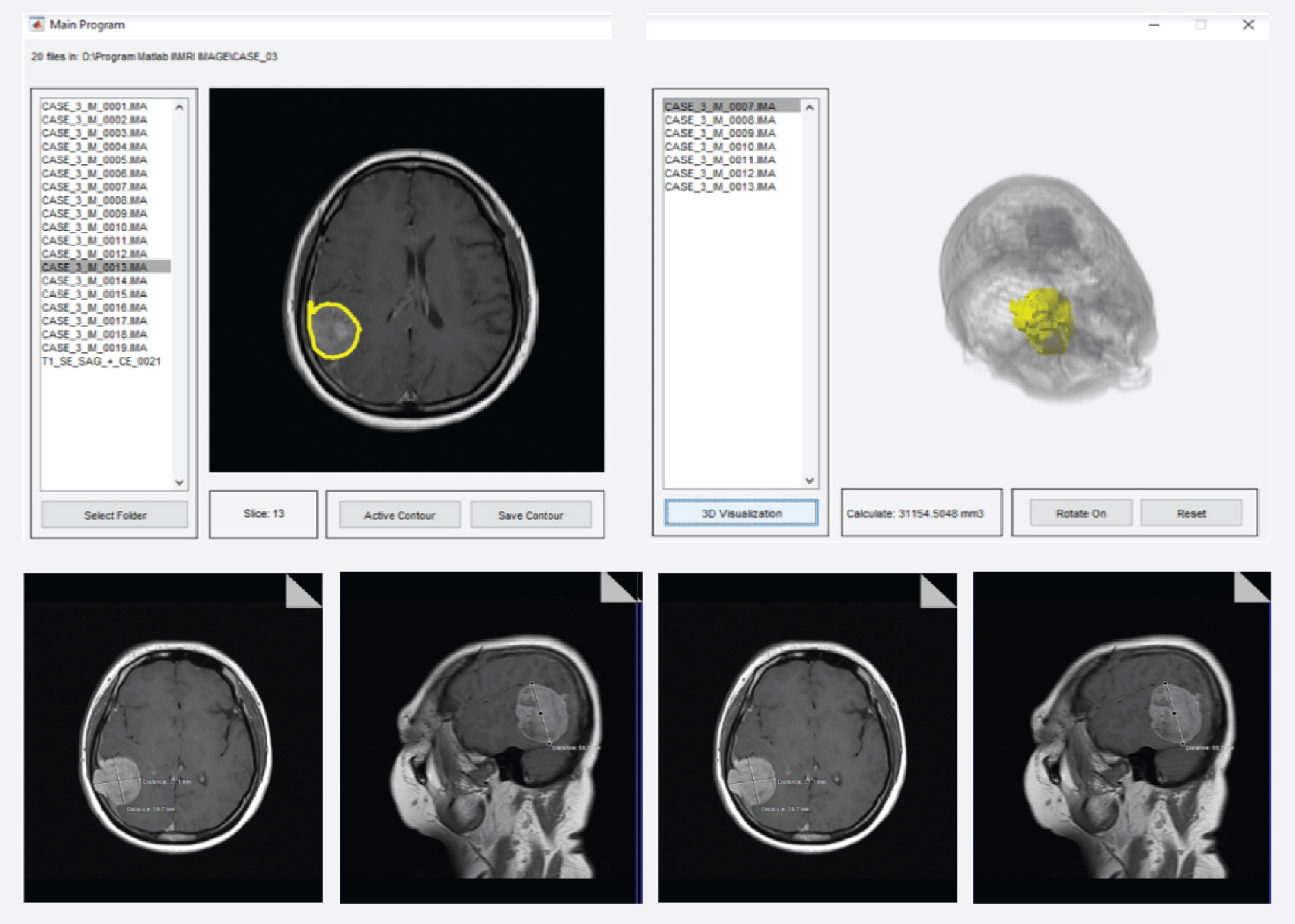
Background: Evaluation of brain tumor MRI image results performed by radiologists employing the linear measurement method has several weaknesses and is sensitive to subjectivity. Purpose: To compare the results of measurements and calculations of brain tumors utilizing the linear measurement method on the Siemens 1.5 Tesla MRI modality employing pulse sequence spin echo with T1 contrast weighting compared with the results of measurements and calculations of brain tumors utilizing the active contour segmentation method. Method: An experimental study was conducted on 32 MRI images. Result: The study's findings indicated that the linear measurement was more significant than the active contour segmentation method (p-value<0,05). The results were obtained by calculating the sensitivity and specificity values of the diagnostic test, which were calculated to be 87.5%. Conclusion: The active contour segmentation method applied to pulse sequence spin-echo T1-weighted contrast can be utilized as an alternative measurement and calculation of brain tumors with a sensitivity and specificity value of 87.5%. Further research suggests developing a Matlab application to compare the results of measurements and calculations of brain tumors on acquiring 3D image magnetic resonance imaging data.
Abdulbaqi, H.S., Jafri, M.Z.M., Mutter, K.N., Omar, A.F., 2016. Segmentation and Estimation of Brain Tumor Volume in Magnetic Resonance Images based on T2-Weighted using Hidden Markov Random Field Algorithm. J. Telecommun. Electron. Comput. Eng. Vol. 8(3), Pp. 9-13.
Adriyanto, O., Agung, H., 2018. Deteksi Tepi untuk Indikasi Tumor Otak Menggunakan Metode Sobel dan Morphological Operations Berdasarkan Citra Magnetic Resonance Imaging. Comput. Eng. Sci. Syst. J. Vol. 3(2), Pp. 179-185.
Astuti, L.W., 2019. Ekstrasi Fitur Citra MRI Otak Menggunakan Data Wavelet Transform (DWT) untuk Klasifikasi Penyakit Tumor Otak. J. Ilm. Inform. Glob. Vol. 10(2), Pp. 80-86.
Bangare, S.L., Patil, M., Bangare, P.S.T., Patil, S., 2015. Implementing Tumor Detection and Area Calculation in MRI Image of Human Brain using Image Processing Techniques. Int. J. Eng. Res. Appl. Vol. 5(4), Pp. 60-65.
Basyid, F., Adi, K., 2014. Segmentasi Citra Medis untuk Pengenalan Objek Kanker Menggunakan Metode Active Contour. Youngster Phys. J. Vol. 3(3), Pp. 209-216.
Currie, S., Hoggard, N., Craven, I.J., Hadjivassiliou, M., Wilkinson, I.D., 2013. Understanding MRI : Basic MR Physics for Physicians. Postgraduated Med. J. Vol. 89(1050), Pp. 209-223.
Jiang, Y., Metz, C.E., 2001. Optimal Method for Combining Two Correlated Diagnostic Assessments with Application to Computer-Aided Diagnosis. In: Medical Imaging (Ed.), Proceedings Image Perception and Performance. Medical Imaging, United States.
Kabade, R.S., Gaikwad, M.S., 2013. Segmentation of Brain Tumour and Its Area Calculation in Brain MR Images using K-mean Clustering and Fuzzy C-Mean Algorithm. Int. J. Comput. Sci. Eng. Technol. Vol. 4(5), 524–531.
Khan, Y., Bhatia, A., 2017. Contour Based Segmentation for Brain Tumor Segmentation in MRI. IJARECE . Vol. 6(11), Pp. 1165–1171.
Kim, H.J., Kim, W., 2012. Method of Tumor Volume Evaluation using Magnetic Resonance Imaging for Outcome Prediction in Cervical Cancer Treated With Concurrent Chemotherapy and Radiotherapy. Radiat. Oncol. J. Vol. 30(2), Pp. 70-77.
Kumar, G.A., Sridevi, P. V., 2019. Active Contour Model for Brain MR Tumor Segmentation snd Volume Estimat. Int. J. Eng. Adv. Technol. Vol. 9(1), Pp. 7226-7231.
Liu, J., Li, M., Wang, J., Wu, F., Liu, T., Pan, Y., 2014. A Survey of MRI-Based Brain Tumor Segmentation Methods. Tsinghua Sci. Technol. Vol. 19(6), Pp. 578-595.
Madhugiri, S.A.S.V.S., Sasidharan, G.M., Kumar, R.V.R., 2016. Measuring Glioma Volumes: A Comparison of Linear Measurement Based Formulae with The Manual Image Segmentation Technique. J. Cancer Res. Ther. Vol. 12(1), Pp. 161-168.
Mbuyamba, E.I., Avina-Cervantes, J.G., Garcia"Perez, A., Romero-Troncoso, R.D.J., 2017. Accepted Manuscript Localized Active Contour Model with Background Intensity Compensation Applied on Automatic MR Brain Tumor Segmentation. Neurocomputing Vol. 220, Pp. 84-97.
Mbuyamba, E.I., Cruz-Duarte, J.M., Avina-Cervantes, J.G., Correa-Cely, C.R., 2016. Active Contours Driven by Cuckoo Search Strategy for Brain Tumour Images Segmentation. Expert Syst. Appl. Vol. 56(1), Pp. 59-68.
Murinto, M., Fitria, R., 2011. Segmentasi Citra Medis MRI (Magnetic Resonance Imaging) menggunakan Segmentasi Citra Medik MRI (Magnetic Resonance Imaging) Menggunakan Metode. In: UAD Yogyakarta (Ed.), Conference: Seminar Nasional Teknik Informatika (STI). UAD Yogyakarta, Yogyakarta, Pp. 1-6.
Priyawati, D., Soesanti, I., Hidayah, I., 2015. Kajian Pustaka Metode Segmentasi Citra pada MRI Tumor Otak. Prosding Semin. Nas. Sains dan Teknol. Fak. Tek. Univ. Wahid Hasyim Vol. 1(1), Pp. 207-215.
R, S.H., K, C., 2013. Tumor Volume Calculation of Brain from MRI Slices. Int. J. Comput. Sci. Eng. Technol. Vol. 4(8), Pp. 1126-1132.
Ro, S., Nag, S., Maitra, I.K., Bandyopadhyay, S.K., 2013. A Review on Automated Brain Tumor Detection and Segmentation from MRI of Brain. Int. J. Adv. Res. Comput. Sci. Softw. Eng. Vol. 3(6), Pp. 1706-1746.
Roy, S., Bandyopadhyay, S., 2012. Detection and Quantification of Brain Tumor from MRI of Brain and it's Symmetric Analysis. Int. J. Inf. Commun. Technol. Res. Vol. 2(6), Pp. 477-483.
Sastroasmoro, S., Ismael, S., 2011. Perkiraan Besar Sampel dalam Penelitian Klinis. In: Dasar-Dasar Metodologi Penelitian. Sagung Seto, Jakarta, Pp. 348.
Siswosudarmo, R., 2017. Tes Diagnostik (Diagnostic Test). Yogyakarta.
Susilo, S., Nagoro, M.T., Kusminarto, K., Budi, W.S., 2011. Uji Diagnostik Pemeriksaan Osteosklerotik Tulang dengan Sistem Radiografi Digital. Media Med. Indones. Vol. 45(3), Pp. 188-193.
Varijki, E., Triwijoyo, B.K., 2017. Segmentasi Citra Mri Menggunakan Deteksi Tepi Untuk Identifikasi Kanker Payudara. Matrik J. Manaj. Tek. Inform. dan Rekayasa Komput. Vol. 15(2), Pp. 17-24.
Westbrook, C., 2014. Handbook of MRI Technique, 2 nd. ed. John Wiley and Sons.
Westbrook, C., Talbot, J., 2019. MRI In Pratice, 5 th. ed. Wiley-Blackwell, United Kingdom.
Widodo, C.E., Adi, K., Sugiharto, A., S., Q.M.B., Pamungkas, A., 2016. Volume Target Delineation for Brain Tumor in MRI Images using Active Contour Segmentation Method. Int. J. Appl. Eng. Res. Vol. 11(16), Pp. 9031-9036.
Widodo, S., 2011. Segmentasi Otomatis untuk Visualisasi 3D Organ Paru pada Citra Computer Tomography menggunakan Active Countour. DUTA.com J. Ilm. Teknol. Inf. da Komun. Vol. 1(2), Pp. 26-40.
Copyright (c) 2022 Journal of Vocational Health Studies

This work is licensed under a Creative Commons Attribution-NonCommercial-ShareAlike 4.0 International License.
- The authors agree to transfer the transfer copyright of the article to the Journal of Vocational Health Studies (JVHS) effective if and when the paper is accepted for publication.
- Legal formal aspect of journal publication accessibility refers to Creative Commons Attribution-NonCommercial-ShareAlike (CC BY-NC-SA), implies that publication can be used for non-commercial purposes in its original form.
- Every publications (printed/electronic) are open access for educational purposes, research, and library. Other that the aims mentioned above, editorial board is not responsible for copyright violation.

Journal of Vocational Health Studies is licensed under a Creative Commons Attribution-NonCommercial-ShareAlike 4.0 International License