SAFIRE STRENGTH OPTIMIZATION: EFFECT ON TISSUE CONTRAST AND PATHOLOGICAL ASSESSMENT OF BRAIN MSCT WITH NON-HEMORRHAGE STROKE (SNH)

Downloads
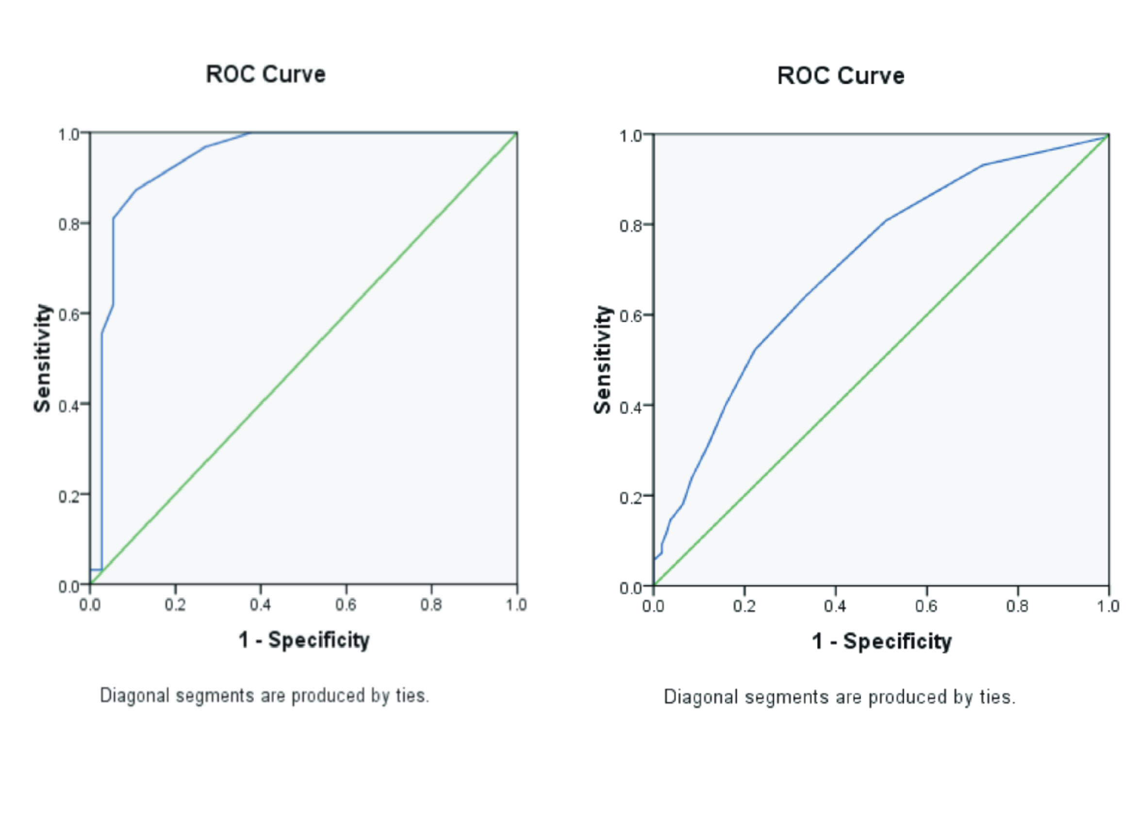
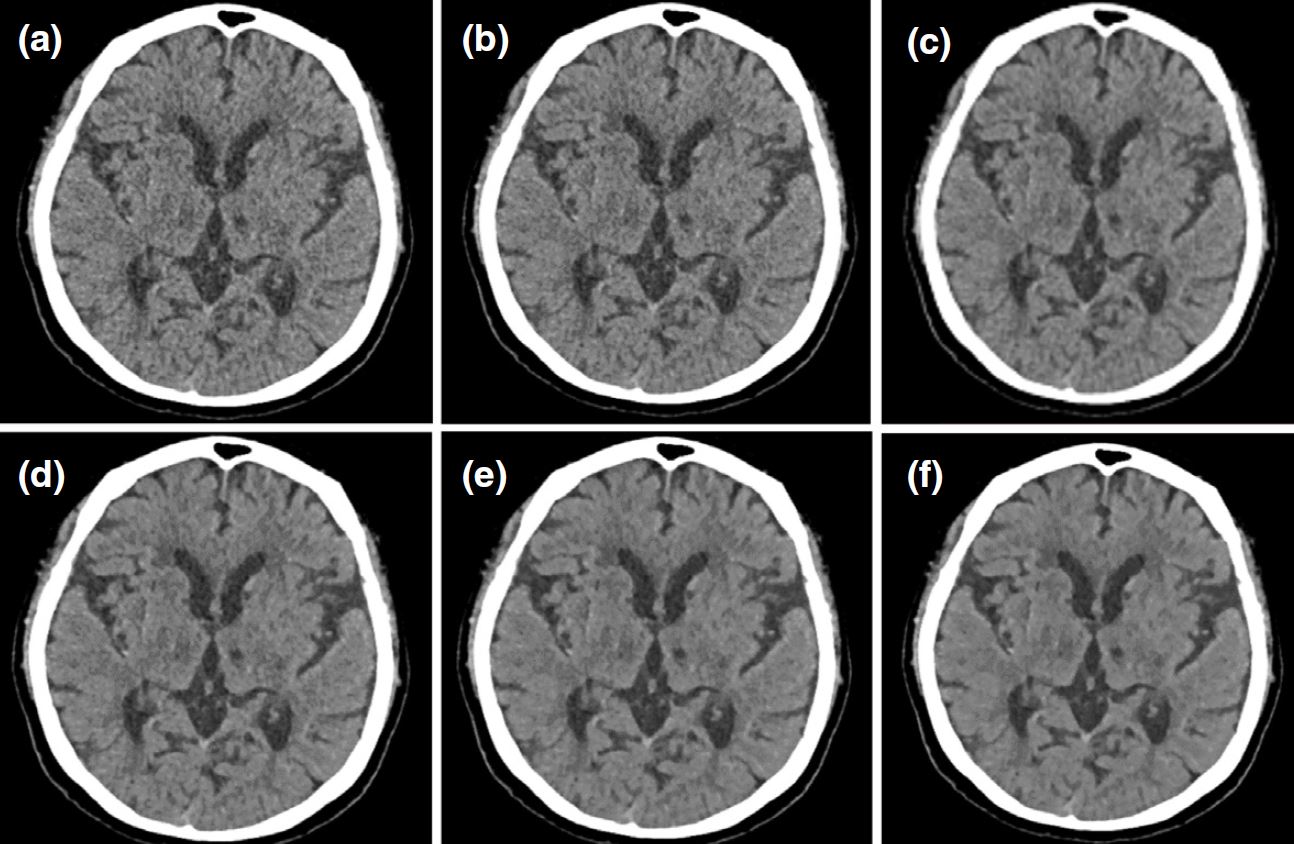
Background: Sinogram Affirmed Iterative Reconstruction (SAFIRE) is an Iterative Reconstruction algorithm that combines IR techniques that utilize raw data and image data iterations as parameters that underlie noise regularization in images in the reconstruction process to improve image quality. Purpose: Analyze the effect of variations in SAFIRE strength values on image contrast and pathological evaluation of CT scan brain with clinical Stroke Non-Hemorrhage (SNH). Method: This research is a quantitative analytic study with an experimental approach to analyze the effect of SAFIRE strength values on image contrast and pathological assessment on CT scan brain examination. Result: Statistical test results showed a significant difference (p-value < 0.05) in all variations of SAFIRE strength, with the resulting Contrast-to-Noise Ratio (CNR) value increasing as the SAFIRE strength value used increased. The average CNR improvement was 18.4% on all SAFIRE strength values compared. This increase is affected by a linear decrease of the noise value from one SAFIRE strength value to another. Image contrast improvement also affects the pathological assessment of SNH due to the increased density differences in the hypodense lesion compared to the surrounding tissues. Conclusion: The use of the SAFIRE strength variation significantly affects image contrast values and pathological assessment in the SNH brain MSCT examination.
Abuelhia, E., Alghamdi, A., 2020. Evaluation Of Arising Exposure of Ionizing Radiation from Computed Tomography and The Associated Health Concerns. Journal of Radiation Research and Applied Sciences. Vol.13(1), Pp. 295–300.
Abuzaid, M.M., Elshami, W., Sulieman, A., Bradley, D., 2022. Cumulative Radiation Exposure, Effective and Organ Dose Estimation from Multiple Head CT Scans in Stroke Patients. Radiation Physics and Chemistry. Vol. 199, Pp. 110306.
Al-Sharify, Z.T., Al-Sharify, T.A., Al-Sharify, N.T., Naser, H.Y., 2020. A Critical Review on Medical ImagingTechniques (CT and PET Scans) in The MedicalField. IOP Conference Series: Materials Science and Engineering. Vol. 870(1). Pp. 012043.
Alsleem, H.A., Almohiy, H.M., 2020. The Feasibility of Contrast-to-Noise Ratio on Measurements to Evaluate CT Image Quality in Terms of Low-Contrast Detailed Detectability. Medical Sciences. Vol. 8(3). Pp. 26.
American College of Radiology, 2020. ACR–ASNR–SPR Practice Parameter For The Performance of Computed Tomography (CT) of The Brain. URL https://www.acr.org/-/media/ACR/Files/Practice-Parameters/ct-brain.pdf (accessed 4.17.23).
Bigdeli, A.H., Nagel, H.D., Antoch, G., Cohnen, M., 2014. Impact of Increasing Levels of Advanced Iterative Reconstruction on Image Quality in Low-Dose Cardiac CT Angiography. Vol. 186 (6). Pp. 567–575.
Bushberg, J. T., Seibert, J. A., Leidholdt, E. M., B.J., 2012. The Essential Physics of Medical Imaging, 3rd Editio. ed. Lippincott Williams & Wilkins, Philadelphia.
Damilakis, J., 2021. CT Dosimetry: What Has Been Achieved and What Remains to Be Done. Investigative Radiology. Vol. 56(1). Pp. 62-68. Pp. 1-10.
DePew, K.D., Boggs, R.C., Yester, M. V., Barnes, G.T., 2022. Direct measurement of CTDIw on helical CT scans. Journal of Applied Clinical Medical Physics. Vol. 23(11). Pp. e13761.
Desai, N., Singh, A., Valentino, D., 2010. Practical Evaluation of Image Quality in Computed Radiographic (CR) Imaging Systems. Proceedings of SPIE - The International Society for Optical Engineering. Vol. 7622. Pp. 1-10.
Feigin, V.L., Brainin, M., Norrving, B., Martins, S., Sacco, R.L., Hacke, W., Fisher, M., Pandian, J., Lindsay, P.,2022. World Stroke Organization (WSO): GlobalStroke Fact Sheet 2022. International Journal ofStroke : Official Journal of The International Stroke Society. Vol. 17(1), Pp. 18–29.
Jung, H., 2021. Basic Physical Principles and Clinical Applications of Computed Tomography. Progress in Medical Physics. Vol. 32(1), Pp. 1–17.
Lee, J.C.Y., 2018. High-pitch Dual-source Computed Tomography Coupled with Sinogram-affirmed Iterative Reconstruction: Image Quality and Radiation Dose in Children. Hong Kong Journal of Radiology. Vol. 21(1), Pp. 40–47.
Mendelson, S.J., Prabhakaran, S., 2021. Diagnosis and Management of Transient Ischemic Attack and Acute Ischemic Stroke: A Review. JAMA. Vol. 325(11), Pp. 1088–1098.
Méndez-Gallardo, J.J., Mendez, B., Cano-Nigenda, V., Farington-Terrero, E.Y., Manrique-Otero, D., Castellanos-Pedroza, E., Merino, J.G., Arauz, A., 2020. Update on The Management of Acute Stroke. A Practical Clinical Guide. Revista Mexicana de Neurociencia. Vol. 21(4). Pp. 163-177.
Ministry of Health, 2018. Kementerian Kesehatan Republik Indonesia. Laporan Nasional RISKESDAS.
Moscariello, A., Takx, R.A.P., Schoepf, U.J., Renker, M., Zwerner, P.L., O'Brien, T.X., Allmendinger, T., Vogt, S., Schmidt, B., Savino, G., Fink, C., Bonomo, L., Henzler, T., 2011. Coronary CT Angiography: Image Quality, Diagnostic Accuracy, and Potential for Radiation Dose Reduction using A Novel Iterative Image Reconstruction Technique-Comparison with Traditional Filtered Back Projection. European Radiology. Vol. 10, Pp. 2130–2138.
Nagayama, Y., Nakaura, T., Tsuji, A., Urata, J., Furusawa, M., Yuki, H., Hirarta, K., Kidoh, M., Oda, S., Utsunomiya, D., Yamashita, Y., 2017. Radiation Dose Reduction using 100-kVp and A Sinogram-Affirmed Iterative Reconstruction Algorithm in Adolescent Head CT: Impact on Grey–White Matter Contrast and Image Noise. European Radiology. Vol. 27(1). Pp. 2717–2725.
Nagayama, Y., Oda, S., Nakaura, T., Tsuji, A., Urata, J., Furusawa, M., Utsunomiya, D., Funama, Y., Kidoh, M., Yamashita, Y., 2018. Radiation Dose Reduction at Pediatric CT: Use of Low Tube Voltage and Iterative Reconstruction. Radiographics. Vol. 38(5), Pp. 1421–1440.
Örgel, A., 2020. Image Quality of CT Angiography of Supra-Aortic Arteries: Comparison Between Advanced Modelled Iterative Reconstruction (ADMIRE), Sinogram Affirmed Iterative Reconstruction (SAFIRE) and Filtered Back Projection (FBP) in One Patients' Group. Clinical Neuroradiology. Vol. 30(1), Pp. 101–107.
Park, J.E., Choi, Y.H., Cheon, J.E., Kim, W.S., Kim, I.O., Cho, H.S., Ryu, Y.J., Kim, Y.J., 2017. Image Quality andRadiation Dose of Brain Computed Tomography inChildren: Effects of Decreasing Tube Voltage from120 kVp to 80 kVp. Pediatric Radiology. Vol. 47(6),Pp. 710–717.
Rahman, M.H., 2020. Radiation Hazard, Safety, Control and Protection. Faridpur Medical College Journa. Vol 14(2), Pp. 100–103.
Retnoningsih & C. Anam, W.S., 2012. Studi Uniformitas Dosis Radiasi CT Scan pada Fantom Kepala yang Terletak pada Sandaran Kepala. Jurnal Sains dan Matematika. Vol. 20(2). Pp. 41-45.
Saini, V., Guada, L., Yavagal, D.R., 2021. Global Epidemiology of Stroke and Access to Acute Ischemic Stroke Interventions. Neurology. Vol. 97 (20 Suppl 2), Pp. S6-S16
Shetewi, S.G., Mutairi, B.S. Al, Bafaraj, S.M., 2020. The Role of Imaging in Examining Neurological Disorders; Assessing Brain, Stroke, and Neurological Disorders using CT and MRI Imaging. Advances in Computed Tomograph. Vol 9, Pp. 1–11.
Singh, R., 2020. Image Quality and Lesion Detection on Deep Learning Reconstruction and Iterative Reconstruction of Submillisievert Chest and Abdominal CT. American Journal of Roentgenology. Vol. 214(3),Pp. 566–573.
Tabrizi, S., Zafar, E., Rafiei, H., 2021. A Cohort Retrospective Study on Computed Tomography Scan Among Pediatric Minor Head Trauma Patients. International Journal of Surgery Open. Vol. 29, Pp. 50–54.
Thibault, J.B., Sauer, K.D., Bouman, C.A., Hsieh, J., 2007. A Three-Dimensional Statistical Approach to Improved Image Quality For Multislice Helical CT. Medical Physics.Vol. 34(11), Pp. 4526–4544.
Tompe, A., Sargar, K., 2021. X-Ray Image Quality Assurance. StatPearls.
Ugwuanyi, D.C., Sibeudu, T.F., Irole, C.P., Ogolodom, M.P., Nwagbara, C.T., Ibekwe, A.M., Mbaba, A.N.,2020. Evaluation of Common Findings in Brain Computerized Tomography (CT) Scan: A SingleCenter Study. AIMS Neuroscience. Vol. 7(3). Pp. 311–318.
Wang, J., 2014. Comparison of Pulmonary Nodule Detection Rate and Accuracy in Low-Dose Chest CT between Iterative Reconstruction Algorithm and Filtered Back Projection Algorithm. Journal of Jilin University Medicine Edition. Vol. 40. Pp. 1098–1103.
Wang, S., 2017. Feasibility of Low Tube Voltage Combined with Iterative Reconstruction in Diagnosis of Urinary Calculus Based on Dual Source CT Dual Energy Scanning Mode. Chinese Journal of Medical Imaging Technology. Vol. 14(1), Pp. 933–936.
Wang, Y., Chen, Y., Fang, J., Song, Y., Shen, J., Wang, J., 2021. Values of Sinogram Affirmed Iterative Reconstruction Algorithm-Based Low-Dose Computed Tomography Imaging in Clinical Diagnosis of Cerebral Hemorrhage. Scientific Programming 2021
Copyright (c) 2024 Journal of Vocational Health Studies

This work is licensed under a Creative Commons Attribution-NonCommercial-ShareAlike 4.0 International License.
- The authors agree to transfer the transfer copyright of the article to the Journal of Vocational Health Studies (JVHS) effective if and when the paper is accepted for publication.
- Legal formal aspect of journal publication accessibility refers to Creative Commons Attribution-NonCommercial-ShareAlike (CC BY-NC-SA), implies that publication can be used for non-commercial purposes in its original form.
- Every publications (printed/electronic) are open access for educational purposes, research, and library. Other that the aims mentioned above, editorial board is not responsible for copyright violation.

Journal of Vocational Health Studies is licensed under a Creative Commons Attribution-NonCommercial-ShareAlike 4.0 International License