HISTOMORPHOLOGICAL ENHANCEMENT OF THE VAGINAL WALL IN DEHYDROEPIANDROSTERONE-TREATED POST-OVARIECTOMIZED WISTAR RATS

Downloads
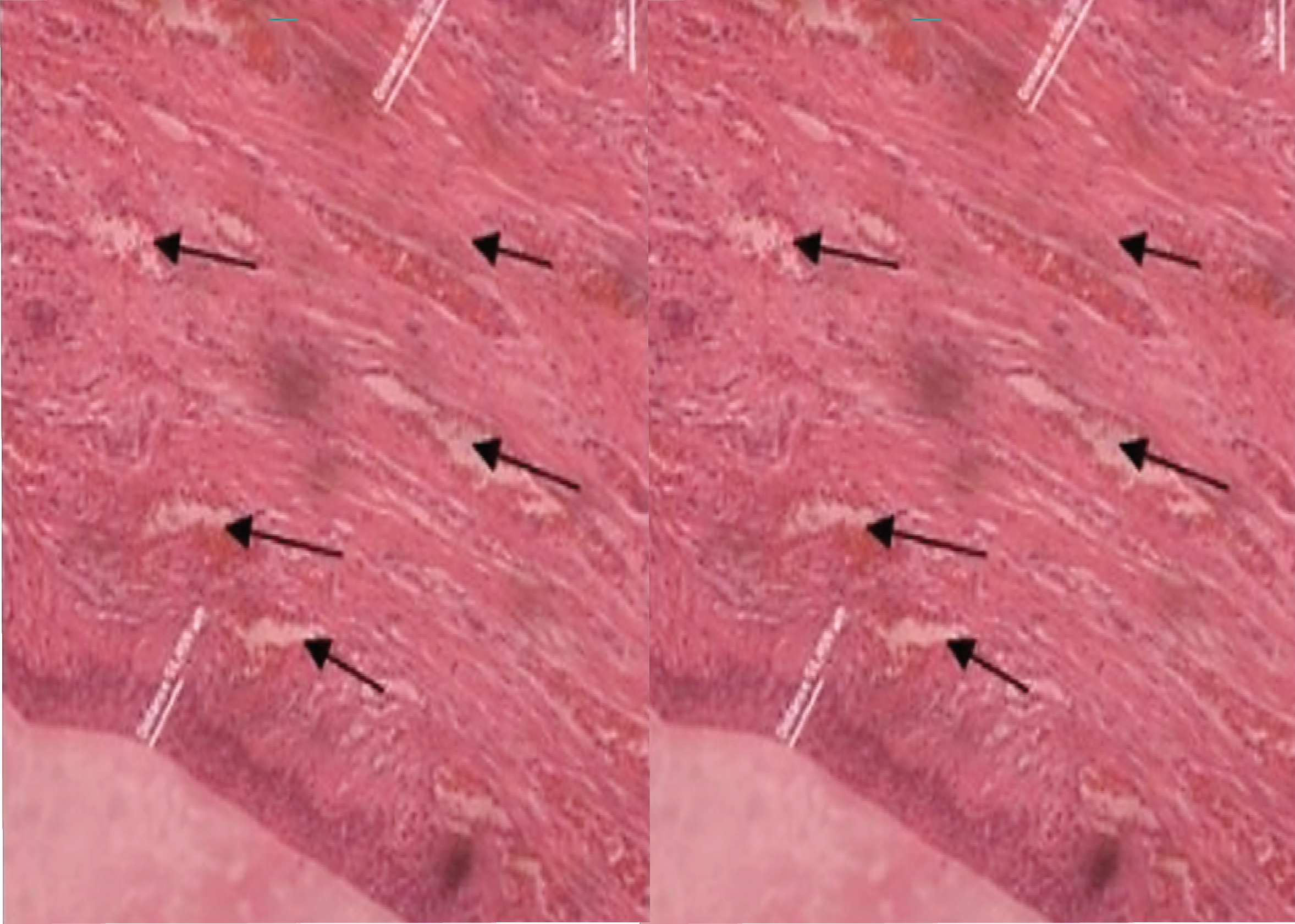
Background: Vaginal atrophy has occurred in three quarters of post-menopausal women. The exclusive source of post-menopausal sex hormones is dehydroepiandrosterone (DHEA). Purpose: Observe the impact of orally administered DHEA in the vagina of a rat (Rattus norvegicus) post- bilateral ovariectomy. Method: This experiment utilized 36 wistar rats aged 10 - 12 weeks with a body weight of 180 - 200 grams. The rats were randomly divided into two groups with an equal number of members. The control group was orally administered glycerin via gavage at a dose of 1 mL per day, while the treatment group received 7.2 mg of DHEA dissolved in glycerin, also administered orally via gavage at a daily dose of up to 1 mL. Both groups were maintained for 42 consecutive days. Finally, a histomorphology examination was conducted on the vaginal tissues of the sacrificed rats. Result: The mean of epithelial and smooth muscle thickness of the treatment group was significantly higher than that of the control group (p-value < 0.05). Besides, the number of blood vessels were also significantly higher in the treated group (p-value < 0.05). Conclusion: Administering DHEA orally via gavage enhances epithelial and smooth muscle tissue thickness, and augments the number of blood vessels in the vagina of wistar rats after bilateral ovariectomy.
Berger, L., El-Alfy, M., Labrie, F., 2008. Effects of Intravaginal Dehydroepiandrosterone on Vaginal Histomorphology, Sex Steroid Receptor Expression and Cell Proliferation in the Rat. J Steroid Biochem Mol Biol Vol. 109(1-2), Pp. 67-80.
Berger, L., El-Alfy, M., Martel, C., Labrie, F., 2005. Effects of Dehydroepiandrosterone, Premarin and Acolbifene on Histomorphology and Sex Steroid Receptors in the Rat Vagina. J Steroid Biochem Mol Biol Vol. 96(2), Pp. 201-215.
Bloch, M., Meiboom, H., Zaig, I., Schreiber, S., Abramov, L., 2013. The Use of Dehydroepiandrosterone in the Treatment of Hypoactive Sexual Desire Disorder: A Report of Gender Differences. European Neuropsychopharmacology Vol. 23(8), Pp. 910-918.
Carlsson, H.E., Hagelin, J., Hagelin, J., 1988. Use of Laboratory Animals in Biomedical and Behavioral Research, in: Use of Laboratory Animals in Biomedical and Behavioral Research. National Academies Press (US).
Davis, S.R., Lambrinoudaki, I., Lumsden, M., Mishra, G.D., Pal, L., Rees, M., Santoro, N., Simoncini, T., 2015. Menopause. Nat Rev Dis Primers Vol. 1(1), Pp. 1-19.
Johnston, S.L., Farrell, S.A., Bouchard, C., Farrell, S.A., Beckerson, L.-A., Comeau, M., Johnston, S.L., Lefebvre, G., Papaioannou, A., SOGC Joint Committee-Clinical Practice Gynaecology and Urogynaecology, 2004. The Detection and Management of Vaginal Atrophy. J Obstet Gynaecol Can Vol. 26(5)], Pp. 503-515.
Jones, M.E., Schoemaker, M.J., Wright, L., McFadden, E., Griffin, J., Thomas, D., Hemming, J., Wright, K., Ashworth, A., Swerdlow, A.J., 2016. Menopausal Hormone Therapy and Breast Cancer: What is the True Size of the Increased Risk? Br J Cancer Vol. 115(5), Pp. 607-615.
Khajuria, D.K., Razdan, R., Mahapatra, D.R., 2012. Description of a New Method of Ovariectomy in Female Rats. Rev Bras Reumatol Vol. 52(3), Pp. 462-470.
Labrie, F., 2019. Intracrinology and Menopause: The Science Describing the Cell-Specific Intracellular Formation of Estrogens and Androgens from DHEA and Their Strictly Local Action and Inactivation in Peripheral Tissues. Menopause Vol. 26(2), Pp. 220-224.
Labrie, F., 2015a. Androgens in Postmenopausal Women: Their Practically Exclusive Intracrine Formation and Inactivation in Peripheral Tissues, in: Rizk, B.R.M.B., Plouffe, J., Leo (Eds.), Androgens in Gynecological Practice. Cambridge University Press, Cambridge, Pp. 64-73.
Labrie, F., 2015b. Intracrinology in Action: Importance of Extragonadal Sex Steroid Biosynthesis and Inactivation in Peripheral Tissues in Both Women and Men. J Steroid Biochem Mol Biol Vol. 145, Pp. 131-132.
Labrie, F., 2015c. All Sex Steroids Are Made Intracellularly in Peripheral Tissues by The Mechanisms of Intracrinology After Menopause. J Steroid Biochem Mol Biol Vol. 145, Pp. 133-138.
Labrie, F., 2010. Important Source of Sex Steroids in Men and Even More in Women. Prog Brain Res Vol. 182, Pp. 97-148.
Labrie, F., Bélanger, A., Pelletier, G., Martel, C., Archer, D.F., Utian, W.H., 2017a. Science of Intracrinology in Postmenopausal Women. Menopause Vol. 24(6), Pp. 702-712.
Labrie, F., Labrie, C., 2013. DHEA and Intracrinology at Menopause, a Positive Choice for Evolution of the Human Species. Climacteric Vol. 16(2), Pp. 205-213.
Labrie, F., Martel, C., Pelletier, G., 2017b. Is Vulvovaginal Atrophy Due to a Lack of Both Estrogens and Androgens? Menopause Vol. 24(4), Pp. 452-461.
Liu, D., Iruthayanathan, M., Homan, L.L., Wang, Yiqiang, Yang, L., Wang, Yao, Dillon, J.S., 2008. Dehydroepiandrosterone Stimulates Endothelial Proliferation and Angiogenesis through Extracellular Signal-Regulated Kinase 1/2-Mediated Mechanisms. Endocrinology Vol. 149(3), Pp. 889-898.
Manson, J.E., Aragaki, A.K., Rossouw, J.E., Anderson, G.L., Prentice, R.L., LaCroix, A.Z., Chlebowski, R.T., Howard, B.V., Thomson, C.A., Margolis, K.L., Lewis, C.E., Stefanick, M.L., Jackson, R.D., Johnson, K.C., Martin, L.W., Shumaker, S.A., Espeland, M.A., Wactawski-Wende, J., WHI Investigators, 2017. Menopausal Hormone Therapy and Long-term All-Cause and Cause-Specific Mortality: The Women's Health Initiative Randomized Trials. JAMA Vol. 318(10), Pp. 927-938.
Manson, J.E., Chlebowski, R.T., Stefanick, M.L., Aragaki, A.K., Rossouw, J.E., Prentice, R.L., Anderson, G., Howard, B.V., Thomson, C.A., LaCroix, A.Z., Wactawski-Wende, J., Jackson, R.D., Limacher, M., Margolis, K.L., Wassertheil-Smoller, S., Beresford, S.A., Cauley, J.A., Eaton, C.B., Gass, M., Hsia, J., Johnson, K.C., Kooperberg, C., Kuller, L.H., Lewis, C.E., Liu, S., Martin, L.W., Ockene, J.K., O'Sullivan, M.J., Powell, L.H., Simon, M.S., Horn, L.V., Vitolins, M.Z., Wallace, R.B., 2013. Menopausal Hormone Therapy and Health Outcomes During the Intervention and Extended Poststopping Phases of the Women's Health Initiative Randomized Trials. JAMA Vol. 310(13), Pp. 1353-1368.
Moiety, F.M., Salem, H.A., Mehanna, R.A., Abdel-Ghany, B.S., 2015. Comparative Study on Induction andEffects of Surgical Menopause in A Female RatModel: A Prospective Case Control Study. Int J ClinExp Med Vol. 8(6), Pp. 9403-9411.
Nappi, R.E., Palacios, S., Panay, N., Particco, M., Krychman, M.L., 2016. Vulvar and Vaginal Atrophy in FourEuropean Countries: Evidence from The EuropeanREVIVE Survey. Climacteric Vol. 19(2), Pp. 188-197.
Naumova, I., Castelo-Branco, C., 2018. Current Treatment Options for Postmenopausal Vaginal Atrophy. Int J Womens Health Vol. 10, Pp. 387-395.
Pangkahila, W.I., 2015. Evidence-Based Anti-Aging Medicine Hormone-Replacement Therapy, 1 st. ed. TLC Publication.
Pelletier, G., Ouellet, J., Martel, C., Labrie, F., 2013. Androgenic Action of Dehydroepiandrosterone (DHEA) on Nerve Density in the Ovariectomized Rat Vagina. J Sex Med Vol. 10(8), Pp. 1908-1914.
Pelletier, G., Ouellet, J., Martel, C., Labrie, F., 2012. Effects of Ovariectomy and Dehydroepiandrosterone (DHEA) on Vaginal Wall Thickness and Innervation. J Sex Med Vol. 9(10), Pp. 2525-2533.
Peni, L.W., Pangkahila, W., 2018. DHEA Supplementation Increases Epithelial Thickness, Plain Muscle Tissue, and Adult Vaginal Blood Vessels (Rattus norvegicus) after Bilateral Post-Ovarectomy. IJAAM (Indonesian Journal of Anti-Aging Medicine).
Pluchino, N., Ninni, F., Stomati, M., Freschi, L., Casarosa, E., Valentino, V., Luisi, S., Genazzani, A.D., Potí¬, E., Genazzani, A.R., 2008. One-Year Therapy with 10 mg/Day DHEA Alone or in Combination with HRT in Postmenopausal Women: Effects on Hormonal Milieu. Maturitas Vol. 59(4), Pp. 93-303.
Portman, D.J., Gass, M.L.S., Vulvovaginal Atrophy Terminology Consensus Conference Panel, 2014. Genitourinary Syndrome of Menopause: New Terminology for Vulvovaginal Atrophy from The International Society for The Study of Women's Sexual Health and The North American Menopause Society. Maturitas Vol. 79(3), Pp. 349-354.
Portman, D.J., Labrie, F., Archer, D.F., Bouchard, C., Cusan, L., Girard, G., Ayotte, N., Koltun, W., Blouin, F., Young, D., Wade, A., Martel, C., Dubé, R., other participating members of VVA Prasterone Group, 2015. Lack of Effect of Intravaginal Dehydroepiandrosterone (DHEA, Prasterone) on The Endometrium in Postmenopausal Women. Menopause Vol. 22(12), Pp. 1289-1295.
Santos, A.C.D., Conley, A.J., Oliveira, M.F. de, Oliveira, G.B., Viana, D.C., Neto, A.C. de A., 2017. Immunolocalization of Steroidogenic Enzymes in The Vaginal Mucous of Galea Spixii during The Estrous Cycle. Reproductive Biology and Endocrinology Vol. 15(1), Pp. 30.
Sripada, R.K., Marx, C.E., King, A.P., Rajaram, N., Garfinkel, S.N., Abelson, J.L., Liberzon, I., 2013. DHEA Enhances Emotion Regulation Neurocircuits and Modulates Memory for Emotional Stimuli. Neuropsychopharmacol Vol. 38(9), Pp. 1798-1807.
Tummala, S., Svec, F., 1999. Correlation between The Administered Dose of DHEA and Serum Levels of DHEA and DHEAS in Human Volunteers: Analysis of Published Data. Clin Biochem Vol. 32(5), Pp. 355-361.
Van Weerden, W.M., Bierings, H.G., van Steenbrugge, G.J., de Jong, F.H., Schröder, F.H., 1992. Adrenal Glands of Mouse and Rat Do Not Synthesize Androgens. Life Sci Vol. 50(12), Pp. 857-861.
Voelker, R., 2017. Relief for Painful Intercourse. JAMA Vol. 317(1), Pp. 18
Copyright (c) 2024 Journal of Vocational Health Studies

This work is licensed under a Creative Commons Attribution-NonCommercial-ShareAlike 4.0 International License.
- The authors agree to transfer the transfer copyright of the article to the Journal of Vocational Health Studies (JVHS) effective if and when the paper is accepted for publication.
- Legal formal aspect of journal publication accessibility refers to Creative Commons Attribution-NonCommercial-ShareAlike (CC BY-NC-SA), implies that publication can be used for non-commercial purposes in its original form.
- Every publications (printed/electronic) are open access for educational purposes, research, and library. Other that the aims mentioned above, editorial board is not responsible for copyright violation.

Journal of Vocational Health Studies is licensed under a Creative Commons Attribution-NonCommercial-ShareAlike 4.0 International License