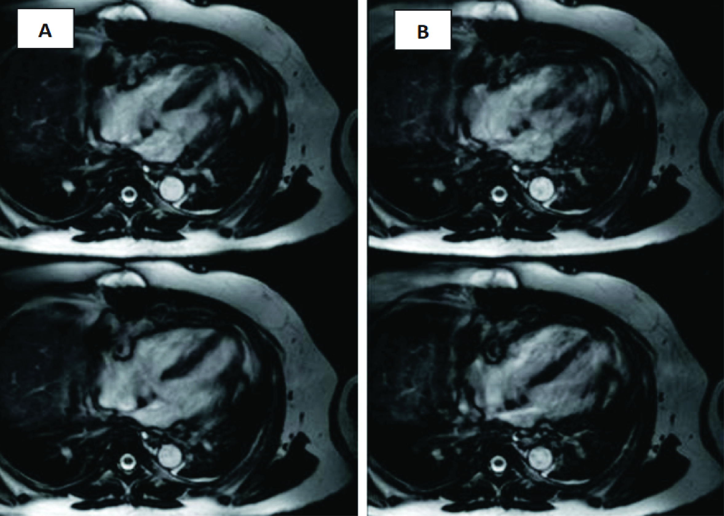
IMAGE QUALITY ANALYSIS 4 CHAMBER SECTIONS OF CARDIAC MRI WITH AND WITHOUT UTILIZING SHIM VOLUME IN THE STEADY STATE FREE PRECESSION SEQUENCES

Downloads
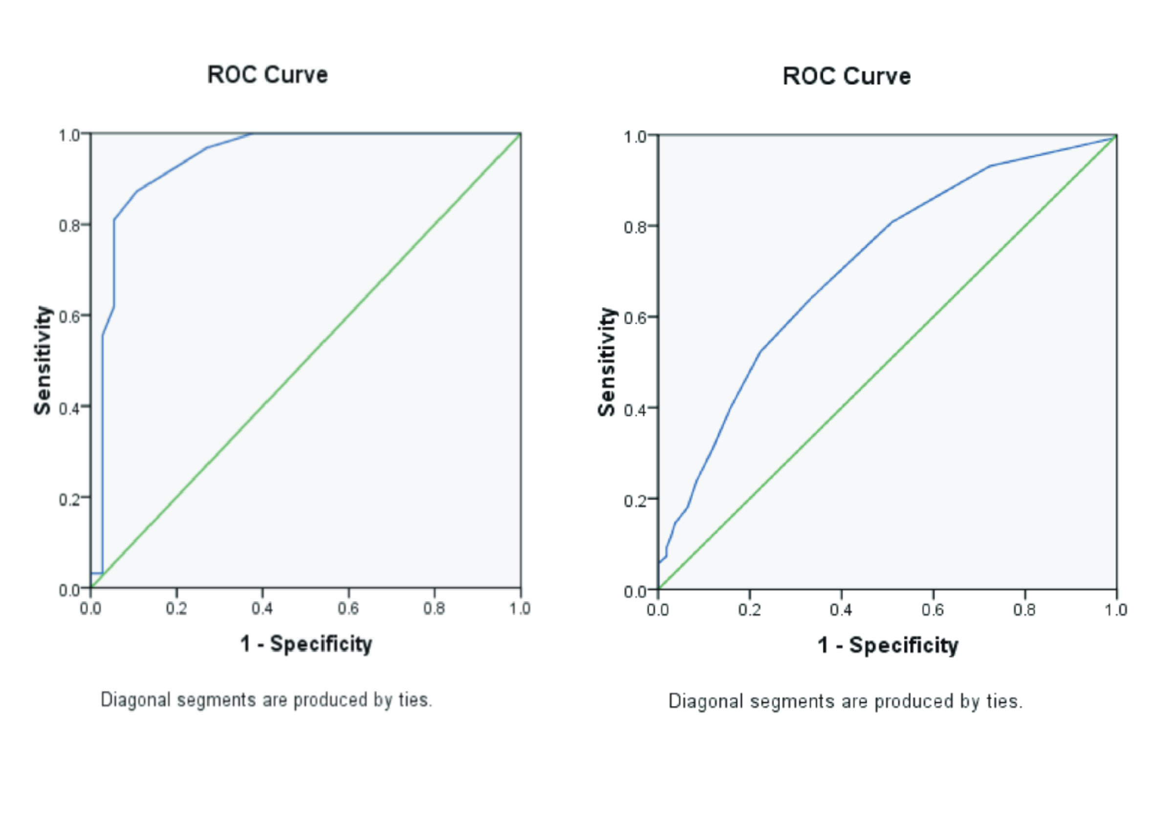
Background: Cardiac MRI examination is relatively rare in Indonesia. The effect of the use of volume shim on moving organs, such as the heart is relatively unknown and noticed by radiographers and cardiologists. Purpose: To analyse the image quality of 4 chamber sections of Cardiac Magnetic Resonance Imaging with and without the use of shim volume on Steady State Free Precession (SSFP) sequences so that it can determine the most optimum 4 chamber images to maintain the diagnostic value by physician. Method: This research is designed through quantitative analytic approach with experiment method. The total samples used are 13 subjects ranging from 7 to 80 years old and 5 respondents and the sample undergone into a series of examinations of Cardiac Magnetic Resonance Imaging. Afterward, the 4 chamber sections in SSFP sequence were given different treatment, namely, with and without the use of shim volume. The result will be assessed by the respondents and then it will be done by non-parametric Wilcoxon Two-Sample Test and parametric Paired Sample T Test. Result: Thereafter, the result of statistic from the respondent assessment is that the quality of anatomy images has p=(0.113) whereas the p=(0.354) for the degree of artefact images and clarifies that the anatomy image quality and the degree of artefact is not too much different. Conclusion: The conclusion of the research is with and without the use of shim volume is not too significant to affect the quality of 4 chamber images.
Brown, M.A., Semelka, R.C. 2003. MRI Basic Principles and Applications. 3rd Ed, Hoboken, New Jersey: John Wiley and Sons, Inc.
Kwong, R.Y. 2008. Cardiovascular Magnetic Resonance Imaging. Totowa, New Jersey: Humana Press.Inc.
Lei, X., Liu, H., Han, Y., Cheng, W., Sun, J., Luo, Y., Yang, D., Dong, Y., Chung, Y. 2017. Reference values of cardiac ventricular structure and function by steady-state free-procession MRI at 3.0T in healthy adult chinese volunteers. J.Magn Reson Imaging. 45(6): Pp. 1684-1692
McRobbie, D.W. 2003. MRI From Picture To Proton 2 nd., Cambridge University Press, New York.
Niitsu, M., Tohno, E., Itai, Y. 2003. Fat Suppression Strategies in Enhanced MR Imaging of the Breast : Comparison of SPIR and water excitation sequences. J Magn Reson Imaging. 18(3):Pp. 310-4.
Notosiswoyo, M., Suswati, S. 2004. Pemanfaatan Magnetic Resonance imaging (MRI) Sebagai Sarana Diagnosa Pasien. Media penelitian dan pengembangan Kesehatan. 8(3).Pp.8–13.
Pearce, E.C., 2009. Anatomi dan Fisiologi untuk Paramedis, Jakarta: PT Gramedia Pustaka Utama.
Phalke, V. V., Gujar, T. Quint, D.J. 2006. Comparison of 3 . 0 T versus 1 . 5 T MR : Imaging of the Spine. Neuroimaging Clin N Am. 16(2):241-8.
Purba, B.A., 2013. Kardiovaskuler. Fakultas Kedokteran dan Ilmu Kesehatan Universitas Jambi.
Reimer, P., Parizel, P.M., Meaney, J.F.M., Stichnoth, F.A. 2010. Clinical MRI Imaging: A Practical Approach. 3rd Ed., Berlin, German: Springer.
Saremi, F., Grizzard, J.D., Kim, R.J., 2008. Optimizing cardiac MR imaging: practical remedies for artifacts. Radiographics : a review publication of the Radiological Society of North America, Inc, 28(4), pp.1161–87.
Sasongko, A., 2015. Adenosine Stress Cardiac Magnetic Resonance An Introduction. In Surabaya,Jawa Timur: Kongres Nasional PARI XIII, p. 28.
Schär, M. Kozerke, S., Fischer, S.E., Boesiger, P. 2004. Cardiac SSFP Imaging at 3 Tesla. Magnetic Resonance in Medicine. 51(4).Pp.799–806.
Tambayong. 2001. Anatomi dan Fisiologi untuk Keperawatan. 1st Ed. Jakarta: ECG.
Thiele, H. Nagel, E., Paetsch, I., Schnackenburg, B., Bornstedt, A., Kouwenhoven, M., Wahl, A., Schuler. G., Fleck, E. 2001. Functional cardiac MR imaging with steady-state free precession (SSFP) significantly improves endocardial border delineation without contrast agents. Journal of Magnetic Resonance Imaging, 14(4), pp.362–367.
Westbrook, C. 2014. Handbook of MRI Technique Fourth. Cambridge, UK: Wiley blackwell.
Westbrook, C., Roth, C.K., Talbot, J. 2011. MRI In Practice. 4th Ed. Blackwell Publishing Ltd.
Woodward, P. 2001. MRI for Technologists. 2nd ., USA: The McGraw-Hill Companies, Inc.
Copyright (c) 2018 Journal Of Vocational Health Studies

This work is licensed under a Creative Commons Attribution-NonCommercial-ShareAlike 4.0 International License.
- The authors agree to transfer the transfer copyright of the article to the Journal of Vocational Health Studies (JVHS) effective if and when the paper is accepted for publication.
- Legal formal aspect of journal publication accessibility refers to Creative Commons Attribution-NonCommercial-ShareAlike (CC BY-NC-SA), implies that publication can be used for non-commercial purposes in its original form.
- Every publications (printed/electronic) are open access for educational purposes, research, and library. Other that the aims mentioned above, editorial board is not responsible for copyright violation.
Journal of Vocational Health Studies is licensed under a Creative Commons Attribution-NonCommercial-ShareAlike 4.0 International License


