FRACTIONAL ANISOTROPY AND MEAN DIFFUSIVITY VALUE IN 2ND GRADE OF DEGENERATIVE CERVICAL CANAL STENOSIS

Downloads
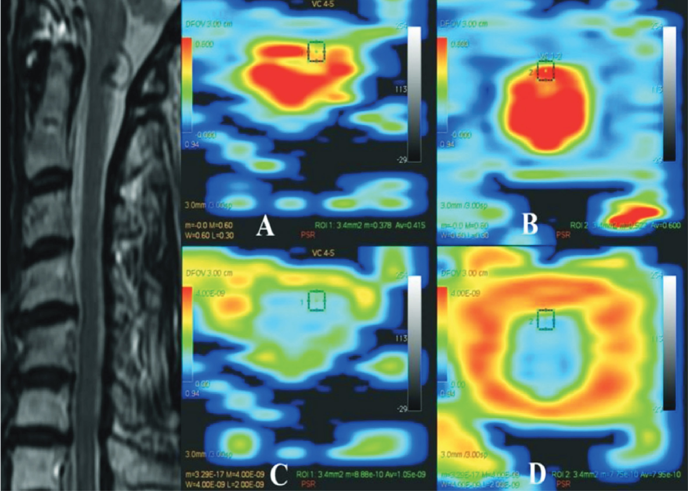
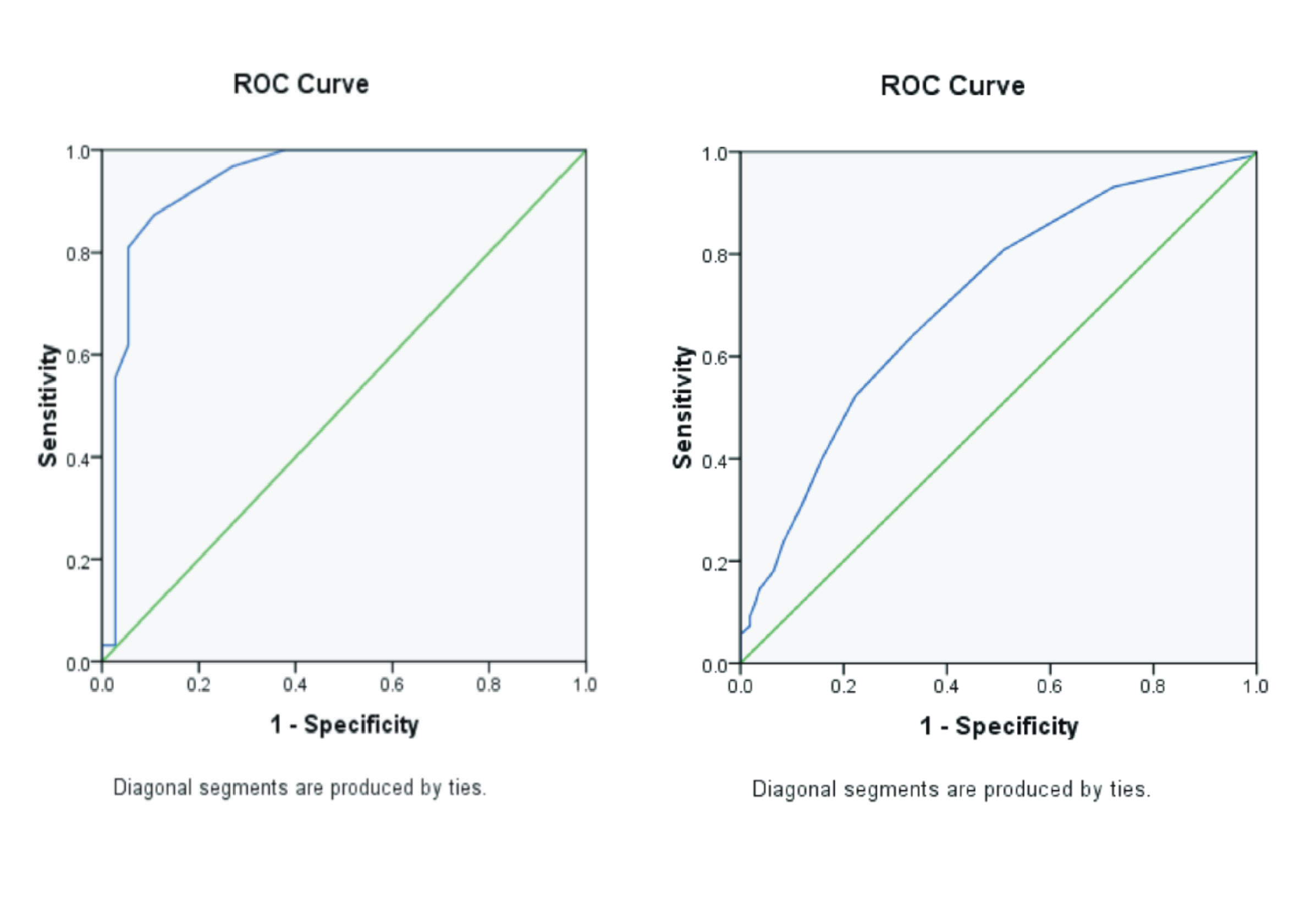
Background: By using T2 weighted image (T2WI) of Magnetic Resonance Imaging (MRI), a radiologist can classify degenerative cervical canal stenosis (DCCS) into three grade, but there is no correlation between stenosis classification with clinical symptoms. It means that radiologist need a new parameter to make an early detection for spinal cord injury (SCI). Purpose: Proving decrease of FA and increase of MD at the most proximal level of 2nd grade DCCS patient compared with C1-2. Method: Cervical MR examination with 15-direction DTI sequens was performed on twenty one patient with neurological signs and symptoms of 2nd grade DCCS. Apparent FA and MD maps were generated on axial plane. The FA and MD measurements in each individual were made at the most proximal level of 2nd grade DCCS and C1-2. Wilcoxon rank sump test was used to compare FA and paired t-test was used for MD. Result : There are significant differences for FA (p = 0,00) and MD (p = 0,00) at the most proximal level of 2nd grade DCCS compared with C1-2. Conclusion: This research shows that FA and MD value at DTI sequens can be used for SCI early detection at 2nd grade DCCS patient
Ahmadli, U., Ulrich, N.H., Yuqiang, Y., Nanz, D., Sarnthein, J., Kollias, S.S. 2015. Early detection of cervical spondylotic myelopathy using diffusion tensor imaging: Experiences in 1.5-tesla magnetic resonance imaging. Neuroradiology Journal Vol. 28(5). Pp 508–514.
Bammer, R., Holdsworth, S.J., Veldhuis, W.B., Skare, S.T. 2009. New Methods in Diffusion-Weighted and Diffusion Tensor Imaging. Magnetic Resonance Imaging Clinics of North America Vol. 17(2). Pp. 175–204.
Fujiyoshi, K., Konomi, T., Yamada, M., Hikishima, K., Tsuji, O., Komaki, Y. 2013. Diffusion tensor imaging and tractography of the spinal cord : From experimental studies to clinical application. Experimental Neurology Vol. 242. Pp. 74–82.
Gulraiz, Quratulain, Farjad, A.S.M. 2017. Chronic Neck Pain and how to Prevent Chronic Neck Pain in Bankers by Using Ergonomic. Journal of Novel Physiotherapies Vol. 7(5). Pp. 1-6.
Kang, Y., Lee, J.W., Koh, Y.H., Hur, S., Kim, S.J., Chai, J.W. 2011. New MRI grading system for the cervical canal stenosis. American Journal of Roentgenology Vol. 197(1). Pp. 134–140.
Kara, B., Celik, A., Karadereler, S., Ulusoy, L., Ganiyusufoglu, K., Onat, L. 2011. The role of DTI in early detection of cervical spondylotic myelopathy: A preliminary study with 3-T MRI. Neuroradiology Journal Vol. 53(8). Pp. 609–616.
Mamata, H., Jolesz, F.A., Maier, S.E. 2005. Apparent Diffusion Coefficient and Fractional Anisotropy in Spinal Cord : Age and Cervical Spondylosis – Related Changes. Journal of Magnetic Resonance Imaging Vol. 22(1). Pp. 38–43.
Northover, J.R., Wild, J.B., Braybrooke, J., Blanco, J. 2012. The epidemiology of cervical spondylotic myelopathy. Skeletal Radiology Vol. 41(12). Pp. 1543–1546.
Rajasekaran, S., Kanna, R.M. 2012. Diffusion tensor imaging of the spinal cord and its clinical applications. The Bone & Joint Journal Vol. 94(8). Pp. 1024–1031.
Shedid, D., Benze,l E.C. 2007. Cervical spondylosis anatomy: Pathophysiology and biomechanics. Neurosurgery Vol. 60(1). Pp. S1-7–S1-13.
Tracy, J.A., Bartleson, J.D. 2010. Cervical Spondylotic Myelopathy. Neurologist Journal Vol. 16(3). Pp. 176–187.
Wheeler-kingshott, C.A.M., Hickman, S.J., Parker, G.J.M., Ciccarelli, O., Symms, M.R., Miller, D.H,. 2002. Investigating Cervical Spinal Cord Structure Using Axial Diffusion Tensor Imaging. NeuroImage Vol. 116(1). Pp. 93–102.
Wu, J-C., Ko, C-C,. Yen, Y-S,, Huang, W-C., Chen, Y-C., Liu ,L. 2013. Epidemiology of Cervical Spondylotic Myelopathy and Its Risk of Causing Spinal Cord Injury: A National Cohort Study. Neurosurg Focus Vol. 35(1):E10.
Young, F.W. 2000. Cervical Spondylotic Myelopathy : A Common Cause of Spinal Cord Dysfunction in Older Persons. American Family Physician Vol. 62(5). Pp. 1064–1070.
Copyright (c) 2019 Journal Of Vocational Health Studies

This work is licensed under a Creative Commons Attribution-NonCommercial-ShareAlike 4.0 International License.
- The authors agree to transfer the transfer copyright of the article to the Journal of Vocational Health Studies (JVHS) effective if and when the paper is accepted for publication.
- Legal formal aspect of journal publication accessibility refers to Creative Commons Attribution-NonCommercial-ShareAlike (CC BY-NC-SA), implies that publication can be used for non-commercial purposes in its original form.
- Every publications (printed/electronic) are open access for educational purposes, research, and library. Other that the aims mentioned above, editorial board is not responsible for copyright violation.
Journal of Vocational Health Studies is licensed under a Creative Commons Attribution-NonCommercial-ShareAlike 4.0 International License


