IMAGE COMPARISON ON T2 QTSE LUMBAL EXAMINATION USING GRAPPA TECHNIQUE WITH AND WITHOUT MAGNETIZATION TRANSFER CONTRAST IN DEGENERATIVE DISC DISEASE CASE

Downloads
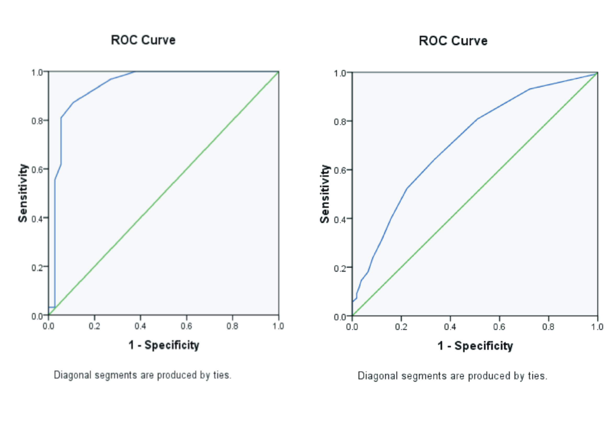
Background: The description of Degenerative Disc Disease in MRI lumbar FSE sequence T2WI is seen as a decrease in signal intensity. Patients with cases of Degenerative Disc Disease experience severe low back pain and cannot lie supine for a long time, while MRI is very sensitive to movement. GRAPPA is a parallel imaging technique that can produce images with a fast scan time but is followed by a decreased Signal to Noise Ratio SNR value. This technique needs to be followed by setting other parameters to produce an optimal image, namely by applying Magnetization Transfer Contrast (MTC). Purpose: To compare the quality of image results on lumbar MRI examination of the sagittal T2 qTSE sequence in the case of Degenerative Disc Disease with and without MTC activation. Method: This research was conducted at the dr. Soedono Madiun from August to September 2020. A sample of 16 patients who met the inclusion criteria was taken during the study. The GRAPPA and GRAPPA+MTC technique imagery results on each sample were assessed for the image quality quantitatively based on the SNR and CNR values. Result: Based on the SNR value, the GRAPPA technique and MTC activation have a higher mean than the GRAPPA technique alone. Likewise, with the CNR value, the GRAPPA technique and MTC activation have a higher average than the GRAPPA technique alone. Conclusion: The GRAPPA technique and MTC activation can be applied in Lumbar MRI examination with cases of Degenerative Disc Disease, especially in uncooperative patients.
Aja-Fernandez, S., Vegas-Sanchez-Ferrero, G., Tristan-Vega, A., 2014. Noise Estimation In Parallel MRI: GRAPPA and SENSE. Magn. Reson. Imaging Vol.32(3), Pp. 281-290.
Apriyani, I., 2019. Perbedaan Kualitas Citra dan Informasi Citra Anatomi Mri Lumbal Sekuen T2 Tse Potongan Sagital dengan dan Tanpa Teknik Parallel Imaging Grappa pada Kasus Hernia Nukleus Pulposus (HNP). Repos. Ris. Kesehat. Nas.
Badan Litbangkes. Politeknik Kesehatan Kemenkes Semarang.
Baruqi, M.S., 2016. Pengaruh Perubahan Time Echo (TE) terhadap Nilai Contrast to Noise Ratio (CNR) Sekuens T2WI TSE Sagital pada Citra MRI Lumbal. Universitas Airlangga.
Boer, R.W., 1995. MR Physics. In: Magnetization Transfer Contrast. Philips Medical Systems, Netherlands, p. Pp. 64-73.
Buller, M., 2018. MRI Degenerative Disease of the Lumbar Spine: A Review. J. Am. Osteopath. Coll. Radiol. Vol.7(4), Pp. 11-19.
Cukke, M.H., Ilyas, M., Murtala, B., Liyadi, F., 2010. Kesesuaian antara Tanda-Tanda Degenerasi Diskus pada Foto Polos Dengan Magnetic Resonance Lumbosakral pada Penderita Nyeri Punggung Bawah.
Donnally, C.J., Hanna, A., Varacallo, M., 2022. Lumbar Degenerative Disk Disease. In: National Library of Medicine. StatPearls Publishing.
Elster, A.D., 2018. How does GRAPPA/ARC work?. MRIquestion.com. URL http://mriquestions.com/grappaarc.html (accessed 2.13.20).
Finelli, D.A., Hurst, G.C., Karaman, B.A., Simon, J.E., Duerk, J.L., Bellon, E.M., 1994. Use of Magnetization Transfer for Improved Contrast on Gradient-Echo MR Images of the Cervical Spine. Radiology Vol.193(1), Pp. 165-171.
Griswold, M.A., Jakob, P.M., Heidemann, R.M., Nittka, M., Jellus, V., Wang, J., Kiefer, B., Haase, A., 2002. Generalized Autocalibrating Partially Parallel Acquisitions (GRAPPA). Magn. Reson. Med. Vol.47(6), Pp. 1202-1210.
Lizak, M.J., Datiles, M.B., Aletras, A.H., Kador, P.F., Balaban, R.S., 2020. MRI of the Human Eye Using Magnetization Transfer Contrast Enhancement. IOVS Investig. Ophthalmol. Vis. Sci. Vol.41(12), Pp. 3878-3881.
Masturoh, I., T., N.A., 2018. Metodologi Penelitian Kesehatan.
Morgan, W.E., Morgan, C.P., 2013. The Lumbar MRI in Clinical Practice: A Survey of Lumbar MRI for Musculoskeletal Clinicians. Independent, Washington DC.
Perez-Torres, C.J., Reynolds, J.O., Pautler, R.G., 2014. Use of Magnetization Transfer Contrast MRI to Detect Early Molecular Pathology in Alzheimer's Disease. Magn. Reson. Med. Vol.71(1), Pp. 333-338.
Pierre, E.Y., Grodzki, D., Aandal, G., Heismann, B., Badve, C., Gulani, V., Sunshine, J.L., Schluchter, M., Liu, K., Griswold, M.A., 2014. Parallel Imaging Based Reduction of Acoustic Noise for Clinical Magnetic Resonance Imaging. Invest. Radiol. Vol.49(9), Pp. 620-626.
Rasad, S., 2005. VII. Toraks -- 6. Tuberkolosis Paru. In: Ekayuda, H.I. (Ed.), Radiologi Diagnostik. Universitas Indonesia, Jakarta, p. Pp. 625.
Ratna, D., 2019. Analisis Citra MRI Sekuen T2 Fluid Attenuation Invers Recovery (FLAIR) dengan Teknik Magnetization Transfer Contrast (MTC) pada Kasus Stroke Iskemik. Universitas Airlangga.
Ruel, L., Brugieres, P., Luciani, A., Breil, S., Mathieu, D., Rahmouni, A., 2004. Comparison of In Vitro and In Vivo MRI of the Spine Using Parallel Imaging. Am. J. Roentgenol. Vol.182(3), Pp. 749-755.
Ryan, M., Cunningham, P., Cantwell, C., Brennan, D., Eustace, S., 2005. A Comparison of Fast MRI of Hips With and Without Parallel Imaging Using SENSE. Br. J. Radiol. Vol.78(928, Pp. 299-302.
Saifudin, S., Hermina, S., Indrati, R., Santjaka, A., 2017. Optimization of R-Factor At GRAPPA Parallel Acquisition Technique on The Image Information T2 Axial Brain MRI. ICASH Vol.1, Pp. 197.
Sayah, A., Jay, A.K., Toaff, J.S., Makariou, E. V, Berkowitz, F., 2016. Effectiveness of a Rapid Lumbar Spine MRI Protocol Using 3D T2-Weighted SPACE Imaging Versus a Standard Protocol for Evaluation of Degenerative Changes of the Lumbar Spine. AJR Am. J. Roentgenol. Vol.3(6), Pp. 614-620.
Suthar, P., Patel, R., Mehta, C., Patel, N., 2015. MRI Evaluation of Lumbar Disc Degenerative Disease. J. Clin. Diagnostic Res. Vol.9(4), Pp. 4-9.
Taher, F., Essig, D., Lebl, D.R., Hughes, A.P., Sama, A.A., Cammisa, F.P., Girardi, F.P., 2012. Lumbar Degenerative Disc Disease: Current and Future Concepts of Diagnosis and Management. Hindawi Publ. Corp. Evidence-Based Complement. Altern. Med. Pp. 1-7.
Westbrook, C., 2014. Handbook of MRI Technique Fourth Edition, 4 th. ed. Wiley-Blackwell, United Kingdom.
Westbrook, C., Roth, C.K., Talbot, J., 2011. MRI In Practice, 4 th. ed. Wiley-Blackwell, United Kingdom.
Weyreuther, M., Heyde, hristoph E., Westphal, M., Zierski, J., Weber, U., Herwig, B., 2007. MRI Atlas Orthopedics and Neurosurgery The Spine, 1 st. ed. Springer, Berlin.
Copyright (c) 2022 Journal of Vocational Health Studies

This work is licensed under a Creative Commons Attribution-NonCommercial-ShareAlike 4.0 International License.
- The authors agree to transfer the transfer copyright of the article to the Journal of Vocational Health Studies (JVHS) effective if and when the paper is accepted for publication.
- Legal formal aspect of journal publication accessibility refers to Creative Commons Attribution-NonCommercial-ShareAlike (CC BY-NC-SA), implies that publication can be used for non-commercial purposes in its original form.
- Every publications (printed/electronic) are open access for educational purposes, research, and library. Other that the aims mentioned above, editorial board is not responsible for copyright violation.
Journal of Vocational Health Studies is licensed under a Creative Commons Attribution-NonCommercial-ShareAlike 4.0 International License


