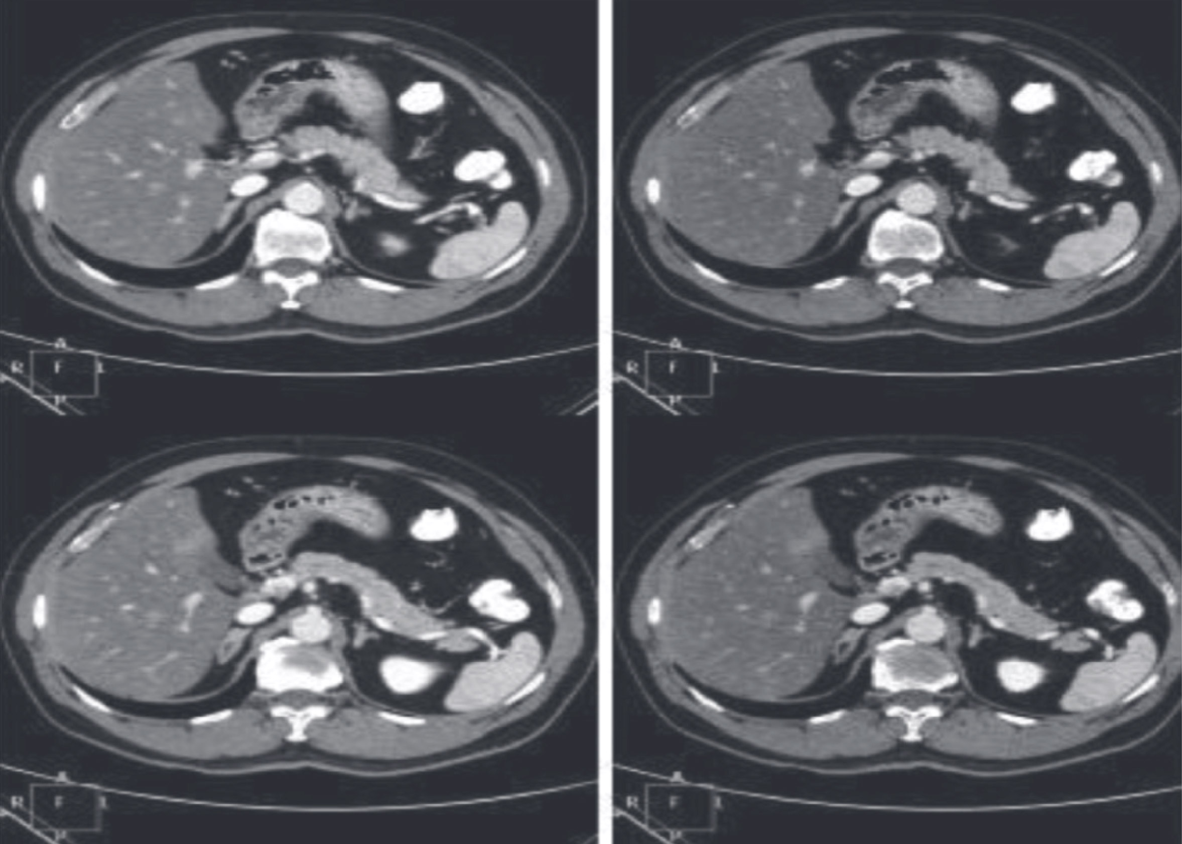
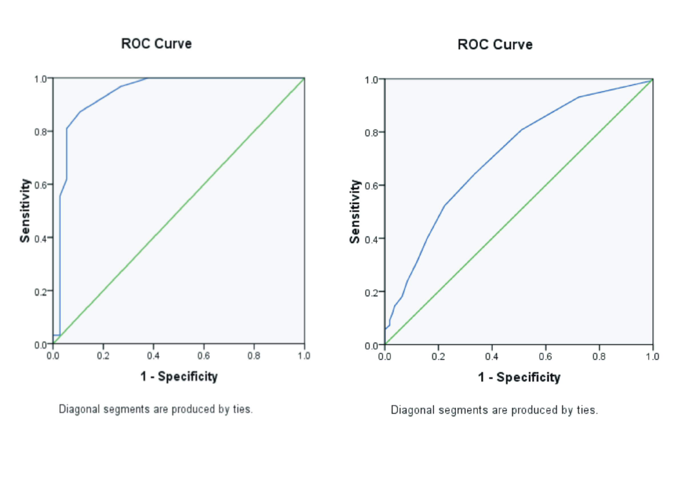
COMPARATIVE ANALYSIS IMAGE RESULTS OF PANCREAS IN PORTAL VEIN PHASE ON CT SCAN ABDOMEN CONTRAST WITHOUT AND WITH MINIMUM INTENSITY PROJECTION IN CT SCAN 64 SLICE

Downloads
Background: Minimum Intensity Projection is a post-proccesing technique on CT Scan that useful for showing structures with low Hounsfiled Unit (HU) values such as pancreas. To demonstrate the anatomy and pathology of the pancreatic organs, a contrast CT scan was performed on pancreatic phase but pancreatic phase was rarely used, so it was replaced by the portal venous phase, but this technique is still rarely used among the radiographers. Purpose: This study aimed to prove the image of the portal venous pancreatic vein on contrast contrast CT scan by using minimum intensity projection (MinIP) on CT scan 64 slices will produce a more optimal image than without the minimum intensity projection (MinIP). Method: This study is a retrospective study with an observational analytic method to assess differences of pancreatic image in contrasting contrast CT scans with and with MinIP reforms on CT 64 slice modalities Philips Briliance. 30 images as samples, with the criteria set by the researchers. The image will be post proccesing without and by using MinIP reformat. Image results will be evaluated by two radiologist, then the data obtained will be tabulated and processed using SPSS software version 17. Result: From this research obtained the result that MinIP reformat able to produce pancreas image more optimal than image without MinIP reformat on CT scan 64 slice and shows a significant difference. Overall assessment of the image has an improvement with the MinIP but for the homogeneity of pancreatic images decreased. Conclusion: There was a significant difference between pancreatic venous porta port results in contrasting CT scans of the abdomen without and with MinIP reformat.
Borghei, P., Shirkhoda, A., Morgan, D. E. 2013. Anomalies, Anatomic Variants, and Sources of Diagnostic Pitfalls in Pancreatic Imaging. Radiology. Vol 266(1). Pp.28-36
Fischer, R., Breidert, M., Keck, T., Makowiec, F., Lohrmann, C., Harder, J. 2012. Early Recurrence of Pancreatic Cancer after Resection and During Adjuvant Chemotherapy. The Saudi Journal of Gastroenterology. Vol 18(2). Pp.118– 122.
Goshima, S., Kanematsu, M., Nishibori, H., Sakurai, K., Miyazawa, D., Watanabe, H., Kondo, H., Shiratori, Y., Onozuka, M., Moriyama, N., Bae, K.T. 2011. CT of the Pancreas : Comparison of Anatomic Structure Depiction, Image Quality, and Radiation Exposure between 320- Detector Volumetric Images and 64- Detector Helical Images.Purpose : Methods : Results ',. Radiology. Vol 260(1). Pp.139–147.
Kim, H.C., Park, S.I., Park, S.J., Shin, H.C. 2004. Diagnosis of Annular Pancreas Using Minimum Intensity Projection of Multidetector Row CT : Case Report. J Korean Radiol Soc. Vol 51. Pp. 641–644.
Koelblinger. 2017. ACR Appropriateness Criteria Staging of Pancreatic Ductal Adenocarcinoma Variant. ACR Appropriateness Criteria.
Qayyum, A., Tamm, E.P., Kamel, I.R., Allen, P.J., Arif-Tiwari, H., Chernyak, V., Gonda, T.A., Grajo, J.R., Hindman, N.M., Horowitz, J.M., Kaur, H., McNamara, M.M., Noto, R.B., Srivastava, P.K., Lalani, T. 2017. ACR Appropriateness Criteria Staging of Pancreatic Ductal Adenocarcinoma Variant. J Am Coll Radiol. 2017. Vol. 14 (11). Pp. 560-569
Lee, E. S., and Lee, J. M. 2014. Imaging diagnosis of pancreatic cancer: A state-of-the-art review. World J Gastroenterol 2014. Vol 20(24). Pp.7864–7877. doi: 10.3748/wjg.v20.i24.7864.
Nakai, M., Sato, M., Ikoma, A., Nakata, K., Saharam S., Takasaka, I., Minamiguchi, H., Kawai, N., Sonomura, T., Kishi, K. 2010. Triple-phase computed tomography during arterial portography with bolus tracking for hepatic tumors. Japanese Journal of Radiology. Vol. 28(2). Pp. 149–156
Quencer, K., Kambadakone, A., Sahani, D., Guimares, A.S.R., 2013. Imaging of the pancreas: Part 1. (online) www.appliedradiology.com.
Salles, A., Nino-murcia, M., Jeffrey, R.B.J. 2008. CT of pancreas : minimum intensity projections. Abdominal Imaging. Vol.33 (2). Pp.207–213.
Vogl, T. J. 2005. Abdominal MDCT : protocols and contrast considerations. Europe Radiology Suppl. Vol 15(5). Pp. 78–90.
Xia, Y., Pan, G., Xue, F., Geng, C. 2013. Reconstruction of the portal vein with 64-slice spiral CT of bile duct obstruction. Experimental And Therapeutic Medicine. Vol 6(101). Pp.401–406.
Copyright (c) 2019 Journal Of Vocational Health Studies

This work is licensed under a Creative Commons Attribution-NonCommercial-ShareAlike 4.0 International License.
- The authors agree to transfer the transfer copyright of the article to the Journal of Vocational Health Studies (JVHS) effective if and when the paper is accepted for publication.
- Legal formal aspect of journal publication accessibility refers to Creative Commons Attribution-NonCommercial-ShareAlike (CC BY-NC-SA), implies that publication can be used for non-commercial purposes in its original form.
- Every publications (printed/electronic) are open access for educational purposes, research, and library. Other that the aims mentioned above, editorial board is not responsible for copyright violation.
Journal of Vocational Health Studies is licensed under a Creative Commons Attribution-NonCommercial-ShareAlike 4.0 International License


