DIFFERENCES VALUE OF PT AND APTT IN EXAMINATION OF ELECTROMECHANICAL AND PHOTO-OPTICAL METHOD

Downloads
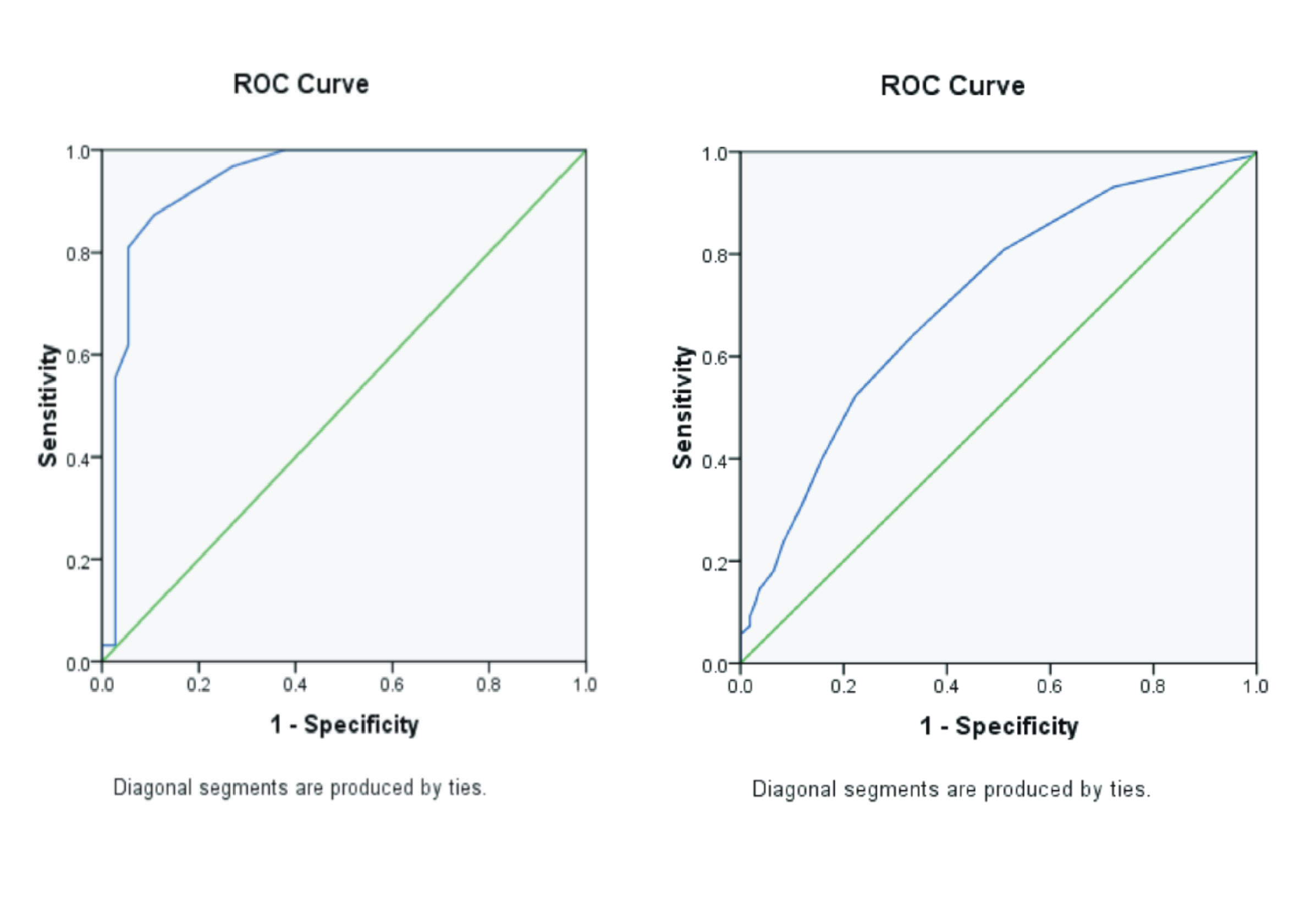
Background: Examination of PT and APTT in hospitals and clinical laboratories by utilizing the use of different instruments and methods. Examination of PT and APTT can be implemented through electromechanical or photo-optical techniques to detect changes in plasma turbidity. This method principle isprincipled that addition can affect the increase in plasma viscosity. Purpose: To analyze the differences in the values of PT and APTT between the electromechanical method and the photo-optical method. Method: Analytical observation, 32 plasma citrate without interference and hemolysis were examined at the Clinical Pathology Laboratory of RSU Haji Surabaya and the Ultra Medica Main Clinics Laboratory in Surabaya. The study used SPSS 24.0 program to determine whether there were PT and APTT values with an electromechanical and photo-optical method. Result: The result of PT and APTT with the electromechanical method was significantly higher than PT and APTT with the photo-optical method on samples without interference. In hemolytic samples, the result of PT with the electromechanical method was significantly higher than the PT result with the photo-optical method. Meanwhile, the result of APTT with the electromechanical method was significantly lower than the APTT result with the photo-optical method in hemolytic samples. Conclusion: There were significant differences in PT and APTT results between electromechanical and photo-optical in samples without interference and hemolytic. It is due to the difference in the detection principle between the two methods.
Aryati, 2014. Peran Patologi Klinik secara Holistik : Tantangan Masa Kini dan Masa Mendatang. In: Kedokteran, F. (Ed.), Pengukuhan Jabatan Guru Besar Dalam Bidang Ilmu Patologi Klinik. Universitas Airlangga, Surabaya, pp. 1–56.
Bakta, I.M., 2007. Hematologi Klinik Ringkas. Buku Kedokteran, EGC, Jakarta.
Castellone, 2011. Interference of Hemolysis, Icteric & Lipemia Coagulation Testing. Elit. Learn.
Durachim, A., Astuti, D., 2018. Bahan Ajar Teknologi Laboratorium Medik (TLM): Hemostasis. Kementrian Kesehatan RI, Jakarta.
Hillman, R.S., Ault, K.A., Leporrier, M., Rinder, H.M., 2011. Hematology in Clinical Practice, 5 th. ed. Mc Graw Hill, New York.
Laga, A.C., Cheves, T.A., Sweeney, J.D., 2006. The Effect of Sample Hemolysis on Coagulation Test Results. Am. J. Clin. Pathol. 126, 748–755.
Lippi, G., Plebani, M., Favaloro, E.J., 2013. Interference in Coagulation Testing: Focus on spurious hemolysis, Icterus and Lipemia. Semin. Thromb. Hemost. 39, 258–266.
Price, S.A., Wilson, L.M.C., 2005. Patofisiologi Klinik Proses-Proses Penyakit. EGC, Jakarta.
Widhiarso, W., 2012. Hasil Uji Statistik dan Penulisan Butir yang Kurang Tepat.
Copyright (c) 2021 Journal of Vocational Health Studies

This work is licensed under a Creative Commons Attribution-NonCommercial-ShareAlike 4.0 International License.
- The authors agree to transfer the transfer copyright of the article to the Journal of Vocational Health Studies (JVHS) effective if and when the paper is accepted for publication.
- Legal formal aspect of journal publication accessibility refers to Creative Commons Attribution-NonCommercial-ShareAlike (CC BY-NC-SA), implies that publication can be used for non-commercial purposes in its original form.
- Every publications (printed/electronic) are open access for educational purposes, research, and library. Other that the aims mentioned above, editorial board is not responsible for copyright violation.
Journal of Vocational Health Studies is licensed under a Creative Commons Attribution-NonCommercial-ShareAlike 4.0 International License


