KLEBSIELLA PNEUMONIAE NECROTIZING FASCIITIS OF THE LOWER EXTREMITY IN A 7-MONTH-OLD MALE: A CASE REPORT
Downloads
Highlights:
- Diagnosing necrotizing fasciitis (NF) was challenging because its symptoms may overlap with other soft tissue infections.
- Necrotizing Fasciitis K. Pneumoniae, a Rare Life-threatening Case.
Abstract:
Introduction: Klebsiella pneumoniae necrotizing fasciitis is an uncommon soft tissue infection characterized by rapidly progressing necrosis involving the skin, subcutaneous tissue, and fascia. This condition may result in gross morbidity and mortality if not treated in its early stages. In fact, the mortality rate of this condition is high, ranging from 25 to 35%. We present a case of 7-month-old male with K. pneumoniae necrotizing fasciitis of the lower extremity.
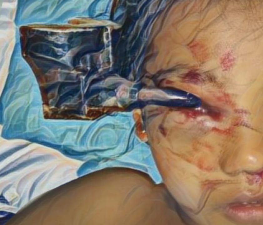
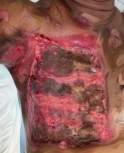
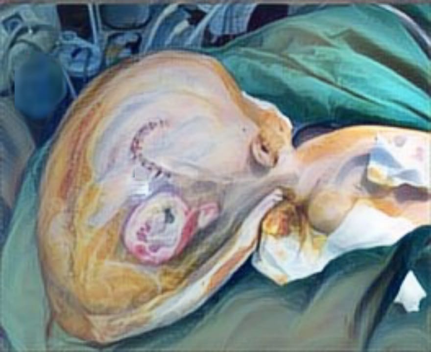
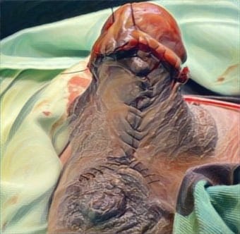
Case Illustration: A 7-month-old male presented with large areas over both left and right inferior side of the lower limbs to the emergency department of Dr. Soetomo General Academic Hospital, Surabaya, Indonesia. Physical examination revealed elevated heart rate of 136 times per minute and increased body temperature of 38oC. The large areas on both lower limbs were darkened, sloughed off, and very tender to palpation. A small area over the right hand was erythematous and sloughed off. Laboratory evaluation demonstrated decreased hemoglobin of 6.2 g/dL and elevated leukocyte of 28,850 g/dL. Blood cultures demonstrated that K. pneumoniae was present.
Discussion: NF is usually hard to diagnose during the initial period. The findings of NF can overlap with other soft tissue infections including cellulitis, abscess or even compartment syndrome. The clinical manifestations of NF start around a week after the initiating event, with induration and edema, followed by 24 to 48 hours later by erythema or purple discoloration and increasing local fever In the next 48 to 72 hours, the skin turns smooth, bright, and serous, or hemorrhagic blisters develop. If unproperly treated, necrosis develops, and by the fifth or sixth day, the lesion turns black with a necrotic crust.
Conclusions: K. pneumoniae necrotizing fasciitis is a rare but life- threatening disease. A high index of suspicion is required for early diagnosis and treatment of this condition
Tso DK, Singh AK. Necrotizing fasciitis of the lower extremity: imaging pearls and pitfalls. Br Inst Radiol. 2018.
Zhao J-C, et al. Necrotizing soft tissue infection: Clinical characteristics and outcomes at a reconstructive center in Jilin Province. BMC Infect Dis. 2017.17(1).
Chaudhry AA, et al. Necrotizing fasciitis and its mimics: What radiologists need to know. Am J Roentgenol. 2015.204(1):128-139.
Fayad L, et al. Musculoskeletal Infection: Role of CT in the Emergency Department Radio-graphics.2007.27(6):1723-1736.
Hayeri MR, et al. Soft-Tissue Infections and Their Imaging Mimics: From Cellulitis to Necrotizing Fasciitis. Radio Graphics. 2016. 36(6): 1888-1910.
Paramythiotis D, et al. Necrotizing soft tissue infections. Surg Pract. 2007; 11(1):17-28.
Puvanendran R et al. Necrotizing fasciitis. Can Fam Physician. 2009.55(10):981-987.
Wong C-H, Wang Y-S. The diagnosis of necrotizing fasciitis. Curr Opin Infect Dis. 2005.18(2):101-106.
Goldstein EJC, et al. Necrotizing Soft-Tissue Infection: Diagnosis and Management. Clin Infect Dis. 2007; 44(5):705-710. doi:10.1086/511638.
Fustes-Morales A, et al. Necrotizing fasciitis: report of 39 pediatric cases. Arch Dermatol. 2002.138(7):893-899.
Fujisawa N, et al. Necrotizing fasciitis caused by Vibrio vulnificus differs from that caused by streptococcal infection. J Infect. 1998;36(3):313-316.
Laupland KB, et al. Invasive group A streptococcal disease in children and association with varicella-zoster virus infection. Ontario Group A Streptococcal Study Group. Pediatrics. 2000.105(5):E60- E60.
Cheng NC, et al. Recent trend of necrotizing fasciitis in taiwan: Focus on monomicrobial Klebsiella pneumoniae necrotizing fasciitis. Clin Infect Dis. 2012.55(7):930-939.
Ou LF, et al. Bacteriology of necrotizing fasciitis: a review of 58 cases. Chung Hua I Hsueh Tsa Chih. 1993.51(4):271-5
Whallett EJ, et al. Necrotising fasciitis of the extremity. J Plast Reconstr Aesthetic Surg. 2010.63(5).
Kelesidis T, Tsiodras S. Postirradiation Klebsiella pneumoniae-associated necrotizing Fasciitis in the Western hemisphere: A rare but life-threatening clinical entity. Am J Med Sci.2009.338(3):217-224.
Decré D, et al. Emerging severe and fatal infections due to Klebsiella pneumoniae in two university hospitals in France. J Clin Microbiol. 2011.49(8):3012-3014.
Gunnarsson GL, et al. Justesen US. Monomicrobial necrotizing fasciitis in a white male caused by hypermucoviscous Klebsiella pneumoniae. J Med Microbiol. 2009.58(11):1519-1521.
Persichino J, et al. Klebsiella pneumoniae necrotizing fasciitis in a Latin American male. J Med Microbiol. 2012.61(11):1614-1616.
Shon AS, et al. Hypervirulent (hypermucoviscous) Klebsiella Pneumoniae: A new and dangerous breed. Virulence. 2013;4(2):107-118.
Giuliano A, et al. Bacteriology of necrotizing fasciitis. Am J Surg. 1977.134(1):52-57. doi:10.1016/0002- 9610(77)90283-5.
Meleney FL. Hemolytic streptococcus gangrene. Arch Surg. 1924.9(2):317-364.
WILSON B. Necrotizing fasciitis. Am Surg. 1952.18(4):416-431.
W.J. R. Necrotizing fasciitis. Ann Surg. 1970.172(6):957-964.
Freeman HP, et al. Necrotizing fasciitis. Am J Surg. 1981.142(3):377-383.
Vijaykumar, et al. Necrotizing fasciitis with chickenpox. Indian J Pediatr. 2003.70:961- 963.
Brisse S, et al. Virulent clones of Klebsiella pneumoniae: Identification and evolutionary scenario based on genomic and phenotypic characterization. PLoS One. 2009.4(3).
Copyright (c) 2019 Marelno Zakanito, Iswinarno Doso Saputro

This work is licensed under a Creative Commons Attribution-ShareAlike 4.0 International License.
JURNAL REKONSTRUKSI DAN ESTETIK by Unair is licensed under a Creative Commons Attribution-ShareAlike 4.0 International License.
- The journal allows the author to hold copyright of the article without restriction
- The journal allows the author(s) to retain publishing rights without restrictions.
- The legal formal aspect of journal publication accessbility refers to Creative Commons Attribution Share-Alike (CC BY-SA)


