Morphological assessment and characterization of uterine caruncles in Bengal goats
Downloads
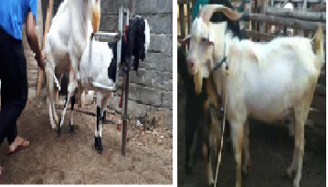
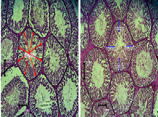
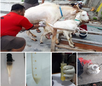
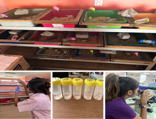
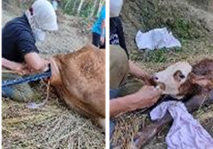
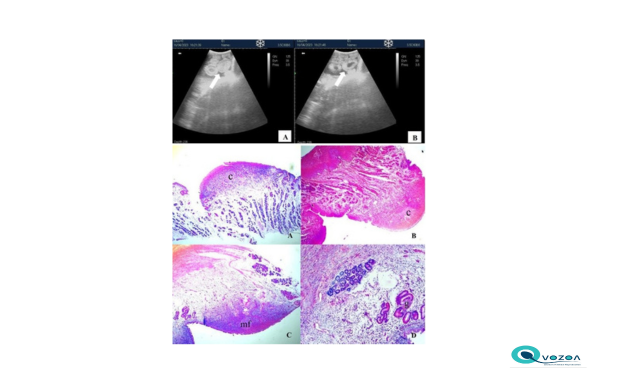
This study aimed to evaluate and characterize the gross, histologic and ultrasonographic features of uterine caruncles in Bengal goats, an indigenous breed in Bangladesh. A total of 40 uteri were collected from sexually mature, pregnant and non-pregnant does obtained from local slaughter houses, preserved and processed in for gross morphological and histological characterization of uterine caruncles. Additionally, transabdominal ultrasonographic characterization of uterine caruncles were performed on randomly selected does at a commercial goat farm. Gross examination revealed multiple dome-shaped caruncles distributed along the endometrial surface, primarily arranged in four longitudinal rows. In both cyclic and non-cyclic goats, the average number of uterine caruncles was higher in the left uterine horn (55.85) compared to the right (54.42), with a consistent average of 4.30 rows observed in both horns. Histological examination of the caruncles revealed dense connective tissue, numerous blood vessels, and abundant uterine glands. The surface epithelium ranged from simple cuboidal to columnar. Morphometric analysis showed that the large caruncles were located in the mid-uterine horns. Transabdominal ultrasonography identified pregnancy in 7 of 20 does (35%) and visualized uterine cotyledons between days 32 and 40 of gestation. Measurement of cotyledons via ultrasound may serve as a reliable indicator of gestational age. These findings provide baseline data that may support reproductive research, enhance breeding management, and contribute to the assessment of reproductive performance and productivity of goats in Bangladesh.
Abd-Elnaeim MMM. 2008. Scanning electron microscopic study on the cyclic goat uterus with special reference to the micro-vascular architecture of the uterine caruncles. J Agric Vet Sci., Qassim Uni. 1: 21-32.
Abdelghafar RM, Bakhiet AO, Ahmed BH. 2007. B-mode real-time ultrasonography for pregnancy diagnosis and fetal number in Saanen goats. J Anim Vet Adv. 6: 702-5.
Abiaezute CN, Nwaogu IC, Okoye CN. 2017. Morphological features of the uterus during postnatal development in the West African Dwarf goat (Capra hircus). Anim Reprod. 14:1062-71.
Adeyinka FD, Laven RA, Lawrence KE, van Den Bosch M, Blankenvoorde G, Parkinson TJ. 2014. Association between placentome size, measured using transrectal ultrasonography, and gestational age in cattle. N Z Vet J 62: 51-6.
Al-Fatlawy HJ. 2015. A comparison of gravid uterine parameters of local breed ewes between single and twin pregnancies in different gestational stages. AL-Qadisiya J Vet Med Sci. 14: 61-4.
Ali MA, Islam MF, Rahman SL, Zohara BF. 2020. Pregnancy diagnosis in goat by using vaginal cytology and trans-abdominal ultrasonography. J Anim Reprod Biotechnol. 35: 338-46.
Amoroso ES. 1952. Placentation. In: Parkes AS (Ed). Marshall's Physiology of Reproduction. 3rd Ed. Longmans Green, London, UK. 127-316..
Anwar M, Riaz A, Rafiq M. 2008. Use of ultrasonography for pregnancy diagnosis in Balkhi sheep. Pak Vet J. 28: 144-6.
Anya KO, Ekere SO, Ogwu DO. 2017. Early pregnancy diagnosis using trans-abdominal ultrasonography in West African dwarf goats. Niger Vet J. 38: 311-8. 10.4314/nvj.v38i4.6.
Atkinson BA, King GJ, Amoroso EC. 1984. Development of the caruncular and intercaruncular regions in the bovine endometrium. Biol Reprod. 30: 763-74.
Bazer FW. 1975. Uterine protein secretions: relationship to development of the conceptus. J Anim Sci. 41: 1376-82.
Budras KD, Habel RE. 2003. Bovine anatomy: an illustrated text. Schlutersche, Hannover, Germany.
Choudhury MP, Sarker SC, Islam F, Ali A, Bhuiyan AK, Ibrahim MN, Okeyo AM. 2012. Morphometry and performance of Black Bengal goats at the rural community level in Bangladesh. Bangladesh J Anim Sci. 41: 83-9.
Cooke PS, Spencer TE, Bartol FF, Hayashi K. 2013. Uterine glands: development, function and experimental model systems. Mol Hum Reprod. 19: 547-58.
DLS. 2023. The livestock economy at a glance. Department of livestock services, BBS 2022-2023.
Doize F, Vaillancourt D, Carabin H, Belanger D. 1997. Determination of gestational age in sheep and goats using transrectal ultrasonographic measurement of placentomes. Theriogenology 48: 449-60.
Dyce KM, Sack WO, Wensing CJG. 2002. Veterinary Anatomy. 3rd Ed. Saunders, Philadelphia, USA. 637- 9.
Ferdous MR, Khan MJ, Rashid MA, Kamruzzaman M. 2011. Effect of different levels of concentrate supplementation on the performance of Black Bengal goat. Bangladesh J Anim Sci. 40: 40-5.
Gray CA, Bazer FW, Spencer TE. 2001. Effects of neonatal progestin exposure on female reproductive tract structure and function in the adult ewe. Biol Reprod. 64: 797-804.
Igwebuike UM. 2009. A review of uterine structural modifications that influence conceptus implantation and development in sheep and goats. Anim Reprod Sci. 112: 1-7.
Igwebuike UM, Ezeasor DN. 2013. The morphology of placentomes and formation of chorionic villous trees in West African Dwarf goats (Capra hircus). Vet Arh. 83: 313-21.
Islam MR, Shamsuddin M, Das PM, Dewan ML. 2001. An outbreak of peste des petits ruminants in Black Bengal goats in Mymensingh, Bangladesh. Bangladesh Vet. 18: 14-9.
Karen A, El Amiri B, Beckers JF, Sulon J, Taverne MA, Szenci O. 2006. Comparison of accuracy of transabdominal ultrasonography, progesterone and pregnancy-associated glycoproteins tests for discrimination between single and multiple pregnancy in sheep. Theriogenology 66: 314-22.
Kumar M, Ray S, Singh KN, Vishen AS, Choudhary PK, Gupta RK. 2020. Histological studies on the ovary and uterus of Black Bengal Goat. J Entomol Zool Stud. 8: 31-3.
Lyngset O. 1971. Studies on reproduction in the goat. Acta Vet Scand. 12: 185-201.
Rasheed YM. 2016. Ultrasonic estimation of gestation age in goats via placentomes diameter. Iraqi J Vet Med. 40: 100-6.
Shehan NA, Kareem DA, Hussein HA, ALkhalad WJ, Jassem ES. 2019. Morphological and histological study of uterus in domestic Iraqi sheep. Al-Qadisiyah J Vet Med Sci. 18: 117-22.
Siddiki AZ, Uddin MB, Hasan MB, Hossain MF, Rahman MM, Das BC, Sarker MS, Hossain MA. 2010. Coproscopic and haematological approaches to determine the prevalence of helminthiasis and protozoan diseases of Red Chittagong cattle (RCC) Breed in Bangladesh. Pak Vet J. 30: 1-6.
Singh G, Prem P. 1990. Effect of age on the morphological changes in the uterus of goat. Indian J Anim Sci. 60: 838-9.
Yotov S, Dimitrov M, Vassilev N. 2005. Early pregnancy diagnosis and examination of embryofoetal growth in the local ovine breed. Bulg J Agric Sci. 11: 119-24.
Zhou Y, Li X, Zhang X, Li M, Luo N, Zhao Y. 2023. Screening of candidate housekeeping genes in uterus caruncle by RNA-Sequence and qPCR analyses in different stages of goat (Capra hircus). Animals (Basel) 13: 1897.
Copyright (c) 2025 Hafsa Hossain, Md. Rashedul Islam, Maksuda Taslima, Mozahidul Islam Tuser, Nurjahan Akter Juli, Al Wasef, Mahfuzul Islam, Jahagir Alam

This work is licensed under a Creative Commons Attribution-ShareAlike 4.0 International License.
Ovozoa by Unair is licensed under a Creative Commons Attribution-ShareAlike 4.0 International License.
1. The journal allows the author to hold the copyright of the article without restrictions.
2. The journal allows the author(s) to retain publishing rights without restrictions
3. The legal formal aspect of journal publication accessibility refers to Creative Commons Attribution Share-Alike (CC BY-SA).
4. The Creative Commons Attribution Share-Alike (CC BY-SA) license allows re-distribution and re-use of a licensed work on the conditions that the creator is appropriately credited and that any derivative work is made available under "the same, similar or a compatible license”. Other than the conditions mentioned above, the editorial board is not responsible for copyright violation.